. 2017 Dec 8:17:118.
doi: 10.1186/s12935-017-0469-8. eCollection 2017.

miR-155-5p modulates malignant behaviors of hepatocellular carcinoma by directly targeting CTHRC1 and indirectly regulating GSK-3β-involved Wnt/β-catenin signaling

Affiliations

miR-155-5p modulates malignant behaviors of hepatocellular carcinoma by directly targeting CTHRC1 and indirectly regulating GSK-3β-involved Wnt/β-catenin signaling

Gang Chen et al. Cancer Cell Int. .

Abstract

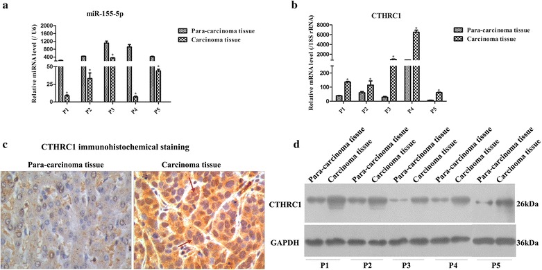
Background: Hepatocellular carcinoma (HCC) remains one of the most lethal cancers. MicroRNA-155 (miR-155) and collagen triple helix repeat containing 1 (CTHRC1) were found to be involved in hepatocarcinogenesis, but their detailed functions in HCC are unclear. Here, we aimed to investigate the underlying role of miR-155-5p and CTHRC1 in HCC.

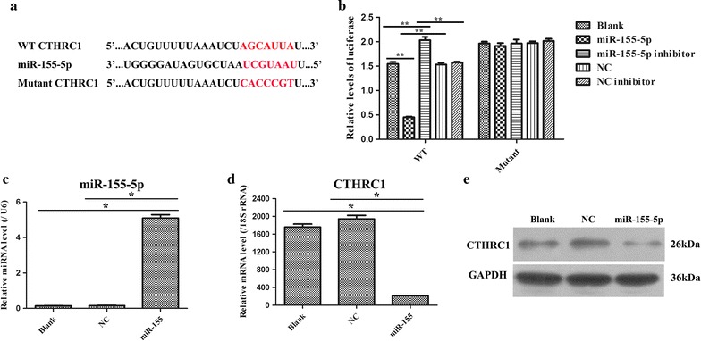
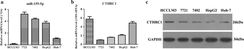
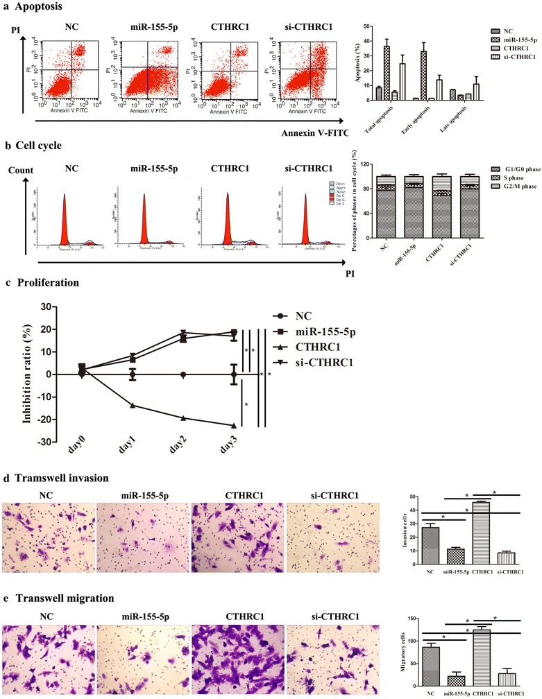
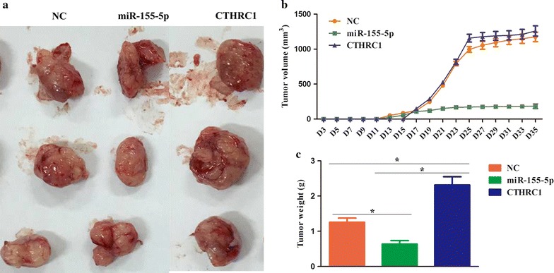
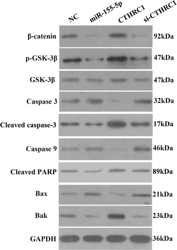
Methods: miR-155-5p and CTHRC1 expression levels were detected by qRT-PCR, IHC and WB in HCC patients and cell lines. Dual-luciferase assay, qRT-PCR and WB were used to validate the target interaction between miR-155-5p and CTHRC1. Biological behaviors, including apoptosis, cell cycle progression, and cell proliferation, invasion and migration, were measured by flow cytometry, CCK-8 assay and Transwell tests. A xenograft model was established to examine the effects of miR-155-5p and CTHRC1 on tumor formation. WB was finally utilized to identify the role of GSK-3β-involved Wnt/β-catenin signaling in HCC growth and metastasis.

Results: Our results showed that miR-155-5p and CTHRC1 were down-regulated and up-regulated, respectively, in HCC patients and cell lines. Dual-luciferase assay verified that CTHRC1 was the direct target of miR-155-5p. Moreover, elevated miR-155-5p expression promoted apoptosis but suppressed cell cycle progression and cell proliferation, invasion and migration in vitro and facilitated tumor formation in vivo; elevated CTHRC1 expression abolished these biological effects. Additionally, miR-155-5p overexpression increased metastasis- and anti-apoptosis-related protein expression and decreased pro-apoptosis-related protein expression, while forced CTHRC1 expression conserved the expression of these proteins.

Conclusion: Altogether, our data suggested that miR-155-5p modulated the malignant behaviors of HCC by targeting CTHRC1 and regulating GSK-3β-involved Wnt/β-catenin signaling; thereby, miR-155-5p and CTHRC1 might be promising therapeutic targets for HCC patients.

Keywords: Collagen triple helix repeat containing 1 (CTHRC1); GSK-3β-involved Wnt/β-catenin signaling; Hepatocellular carcinoma (HCC); Malignant behaviors; microRNA-155-5p (miR-155-5p).


Figures

Fig. 1
Fig. 1
Expression of miR-155-5p and CTHRC1 in the para-carcinoma tissue and carcinoma tissue of HCC patients. a miR-155-5p expression was analyzed by qRT-PCR in the para-carcinoma tissue and carcinoma tissue of HCC patients. miR-155-5p was significantly down-regulated in carcinoma tissue. “p” indicates patient. *p < 0.05. b CTHRC1 mRNA expression was detected by qRT-PCR in the para-carcinoma tissue and carcinoma tissue of HCC patients. CTHRC1 was remarkably up-regulated in carcinoma tissue. *p < 0.05. c Immunohistochemical staining of CTHRC1 in the para-carcinoma tissue and carcinoma tissue of HCC patients (× 200 magnification). The red arrow represents positive staining of CTHRC1, which was mainly located in cytoplasm. d CTHRC1 protein expression was tested by WB in the para-carcinoma tissue and carcinoma tissue of HCC patients. CTHRC1 was notably elevated in carcinoma tissue. *p < 0.05
Fig. 2
Fig. 2
CTHRC1 is a direct target of miR-155-5p. a A graphical representation of miR-155-5p -binding sites in the wild-type CTHRC1-3′-UTR and mutant CTHRC1-3′-UTR. b Dual-luciferase reporter assay was performed in 293T cells, and the data are presented as the mean ± SD. **p < 0.01. c miR-155-5p expression was examined by qRT-PCR in 293T cells after transfection with NC and miR-155-5p mimic plasmids. *p < 0.05. d CTHRC1 mRNA expression was determined by qRT-PCR in 293T cells after transfecting with NC and miR-155-5p mimic plasmids. *p < 0.05. e Western blot analysis of CTHRC1 protein levels in the blank, NC and miR-155-5p mimic groups
Fig. 3
Fig. 3
Expressions of miR-155-5p and CTHRC1 in five strains of HCC cell lines. a The different expression levels of miR-155-5p were analyzed by qRT-PCR in five strains of HCC cell lines. b The different gene expressions of CTHRC1 were measured by qRT-PCR in five strains of HCC cell lines. c The different protein expressions of CTHRC1 were tested by qRT-PCR in five strains of HCC cell lines
Fig. 4
Fig. 4
Assessment of the effect of miR-155-5p on apoptosis, cell cycle, proliferation, cell invasion and cell migration by CTHRC1 in HCCLM3 cells. a miR-155-5p triggered apoptosis via CTHRC1 in HCCLM3 cells. FACS plots in the left panel show the pattern of apoptosis in representative samples from each group. The statistical histogram is on the right panel. b miR-155-5p induced G0/G1 phase cell cycle arrest via CTHRC1 in HCCLM3 cells. Representative cell cycle profiles of each group are presented in the left panel. c miR-155-5p promoted cell proliferation via CTHRC1 in HCCLM3 cells. d miR-155-5p inhibited cell invasion via CTHRC1 in HCCLM3 cells. Representative images of each group in the Transwell invasion assay. Bar charts show the cell invasion ratio in the right panel. *p < 0.05. Scale bars = 100 μm. e miR-155-5p suppressed cell migration via CTHRC1 in HCCLM3 cells. Representative images of each group in the Transwell migration assay. Bar charts show the cell migration ratio in the right panel. *p < 0.05, scale bars = 100 μm
Fig. 5
Fig. 5
miR-155-5p suppressed tumor growth through CTHRC1 in a nude mouse model. a Representative images of tumors in different groups. b Growth curve of tumor volume from 3 to 35 days. c The tumor weights of each group. * p < 0.05
Fig. 6
Fig. 6
miR-155-5p and CTHRC1 mediated the expression of the proteins β-catenin, p-GSK-3β, GSK-3β, Caspase3, Cleaved caspase3, Caspase9, Cleaved PARP, Bax and Bak as determined by WB assay

References

    1. Dominguez-Malagon H, Gaytan-Graham S. Hepatocellular carcinoma: an update. Ultrastruct Pathol. 2001;25:497–516. doi: 10.1080/019131201753343539. - DOI - PubMed
    1. Marotta F, Vangieri B, Cecere A, Gattoni A. The pathogenesis of hepatocellular carcinoma is multifactorial event. Novel immunological treatment in prospect. La Clin Ter. 2004;155:187–199. - PubMed
    1. Coleman WB. Mechanisms of human hepatocarcinogenesis. Curr Mol Med. 2003;3:573–588. doi: 10.2174/1566524033479546. - DOI - PubMed
    1. Shin JW, Chung YH. Molecular targeted therapy for hepatocellular carcinoma: current and future. World J Gastroenterol. 2013;19:6144–6155. doi: 10.3748/wjg.v19.i37.6144. - DOI - PMC - PubMed
    1. Han LL, Lv Y, Guo H, Ruan ZP, Nan KJ. Implications of biomarkers in human hepatocellular carcinoma pathogenesis and therapy. World J Gastroenterol. 2014;20:10249–10261. doi: 10.3748/wjg.v20.i30.10249. - DOI - PMC - PubMed