Review
. 2017 Dec 6:3:28.
doi: 10.1186/s40748-017-0068-7. eCollection 2017.

Nuchal cord and its implications

Affiliations
Review

Nuchal cord and its implications

Morarji Peesay. Matern Health Neonatol Perinatol. .

Abstract

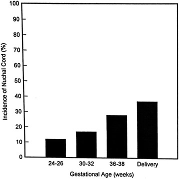
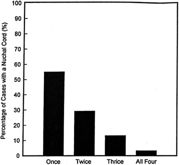
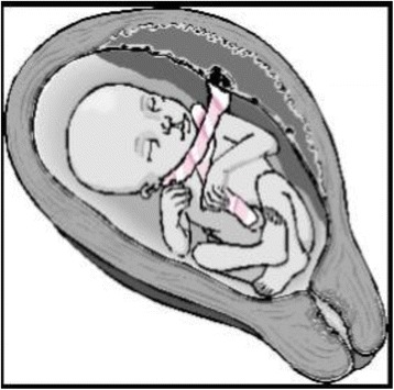
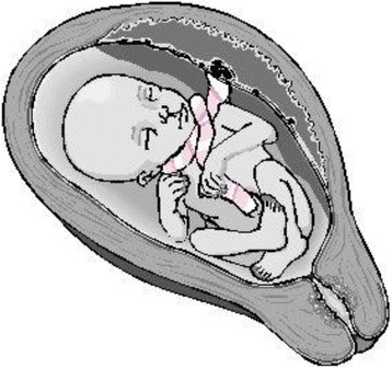
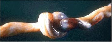
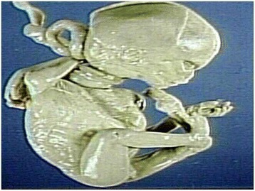
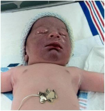
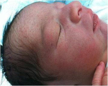
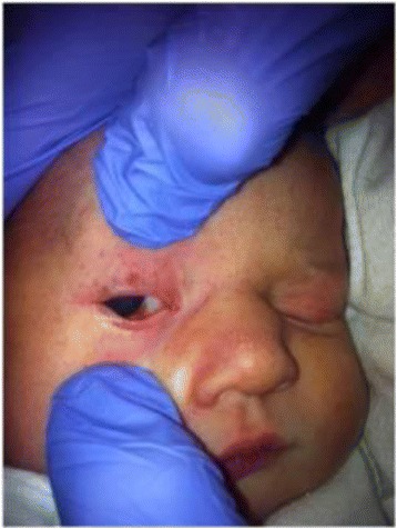
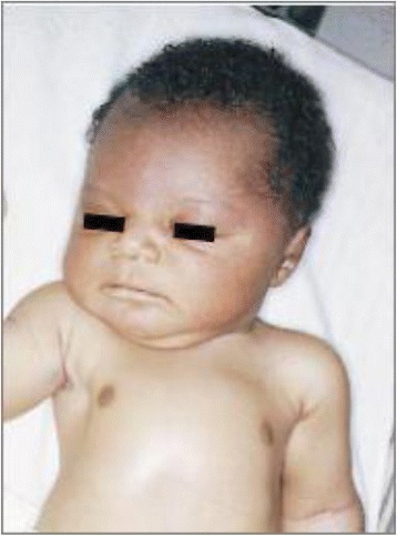
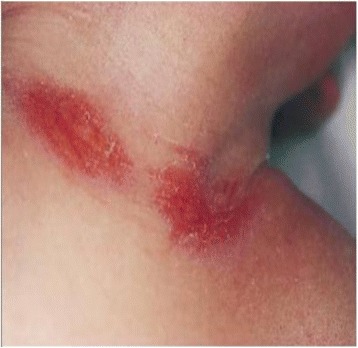
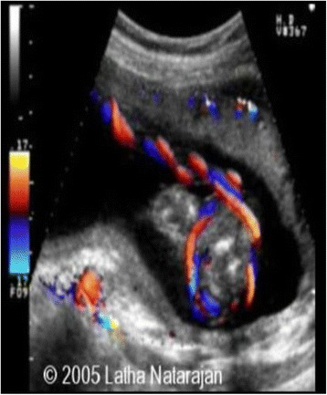
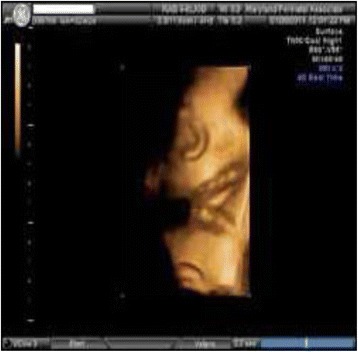
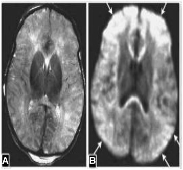
Nuchal cord occurs when the umbilical cord becomes wrapped around the fetal neck 360 degrees. Nuchal cords occur in about 10-29% of fetuses and the incidence increases with advancing gestation age. Most are not associated with perinatal morbidity and mortality, but a few studies have shown that nuchal cord can affect the outcome of delivery with possible long-term effects on the infants. Nuchal cords are more likely to cause problems when the cord is tightly wrapped around the neck, with effects of a tight nuchal cord conceptually similar to strangulation. Umbilical cord compression due to tight nuchal cords may cause obstruction of blood flow in thin walled umbilical vein, while infant's blood continues to be pumped out of the baby through the thicker walled umbilical arteries causing hypovolemia, acidosis and anemia. Some of these infants have physical features secondary to tight nuchal cords that are distinct from those seen with birth asphyxia. The purpose of this article is to review the pathophysiology of tight nuchal cords and explore gaps in knowledge and research areas.

Keywords: Adult strangulation; Nuchal cords; Partial prolonged asphyxia; Tight Nuchal cord around the neck syndrome (tCAN syndrome).


Conflict of interest statement

Ethics approval and consent to participate

Not Applicable.

Consent for publication

All figures and Tables are obtained written consent.

Competing interests

The authors declare that they have no competing interests.

Publisher’s Note

Springer Nature remains neutral with regard to jurisdictional claims in published maps and institutional affiliations.

Figures

Fig. 1
Fig. 1
The incidence of nuchal cords at the four time points studied (Courtesy from Elsevier, with permission)
Fig. 2
Fig. 2
The frequency of occurrence in individual fetuses who had nuchal cord indentified during the four evaluations (Courtesy from Elsevier, with permission)
Fig. 3
Fig. 3
Nuchal Cord - Free sliding pattern: (Courtesy Philippe Jeanty et al., with permission)
Fig. 4
Fig. 4
Nuchal Cord - Locked pattern (Courtesy from Philippe Jeanty et al., with permission)
Fig. 5
Fig. 5
Umbilical Cord Knot. Notice the area of congestion to the right. The congested side is always the side between the knot and the placenta (Courtesy from University of Mo. Department of Pathology, with permission)
Fig. 6
Fig. 6
Pathology specimen of fetus with multiple nuchal cords (Courtesy from Peter Anderson PEIR Digital Library, with permission)
Fig. 7
Fig. 7
Facial Duskiness due to tight Nuchal cord
Fig. 8
Fig. 8
Petechiae on face due to tight nuchal cord
Fig. 9
Fig. 9
Facial Petechiae and Subconjuntival hemorrhage due to tight Nuchal cord
Fig. 10
Fig. 10
Suffusion of the face and head due to tight nuchal cord. (From Arnold J. Rudolph, M.D. Atlas of the Newborn, B.C Decker Inc., with permission)
Fig. 11
Fig. 11
Nuchal Cord with Skin abrasion of neck - Cord Around the Neck times three. (From Arnold J. Rudolph, M.D. Atlas of the Newborn, B.C Decker Inc., with permission)
Fig. 12
Fig. 12
Fetal Ultra sound color doppler scan of Nuchal Cord (Courtesy from Dr. Latha Natarajan, with permission)
Fig. 13
Fig. 13
Fetal Ultra Sound scan - 3D image of double nuchal cord (Courtesy from Maryland Perinatal Associates, with permission)
Fig. 14
Fig. 14
MRI scans of severe Partial Prolonged Asphyxia: A 4-day-old infant owing to tight cord around neck. A) Axial T2- weighted imaging shows abnormal cortical and subcortical bilateral cerebral increased T2 signal intensity. B) Axial diffusion weighted imaging shows extensive gray and white matter injury (arrows) of both cerebral hemispheres – From KN V: Cerebral Palsy and Early Stimulation. New Delhi, India: Jaypee Brothers. Medical Publishers (P) Ltd., 2014, with permission

References

    1. Gould GM, Pyle WL. Prenatal anomalies. Anomalies and curiosities of medicine. New York: The Julian Press, Inc.; 1896. p. 95.
    1. Crawford S. Cord round the neck. Incidence and squeal. Acta Pediátrica. 1962;51:594–603. doi: 10.1111/j.1651-2227.1962.tb06586.x. - DOI - PubMed
    1. Larson JD, Rayburn WF, Crosby S, Thurnau GR. Multiple nuchal cord entanglements and intrapartum complications. Am J Obstet Gynecol. 1995;173:1228. doi: 10.1016/0002-9378(95)91359-9. - DOI - PubMed
    1. Clapp JF, Stepanchak W, Hashimoto K, Ehrenberg H, Lopez B. The natural history of antenatal nuchal cords. Am J Obstet Gynecol. 2003;189:488–493. doi: 10.1067/S0002-9378(03)00371-5. - DOI - PubMed
    1. Henry E, Andres RL, Christensen RD. Neonatal outcomes following a tight nuchal cord. J Perinatol. 2013;33:231. doi: 10.1038/jp.2012.79. - DOI - PubMed

LinkOut - more resources