

Review
doi: 10.1016/j.ijwd.2017.08.001.
eCollection 2017 Dec.
Periocular dermatoses
Affiliations
- PMID: 29234715
- PMCID: PMC5715233
- DOI: 10.1016/j.ijwd.2017.08.001
Item in Clipboard
Review
Periocular dermatoses
Int J Womens Dermatol.
.
Abstract
The periocular area may be affected by infectious or noninfectious diseases such as inflammatory dermatoses, systemic disease, drug reactions, benign and malignant lesions, traumatic lesions, and esthetic complications. We present a review of the most common periocular dermatoses.
Figures
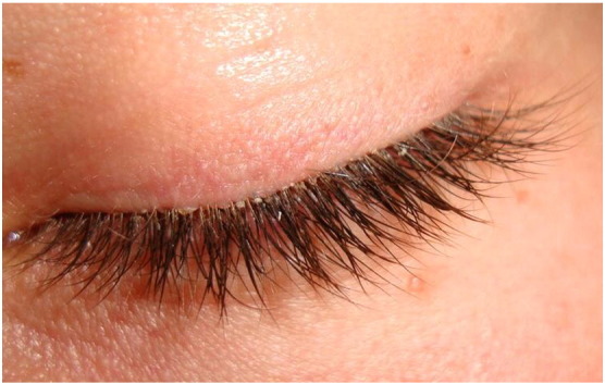
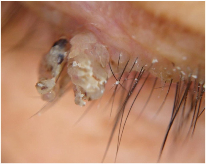
Demodecidosis that causes chronic blepharitis.
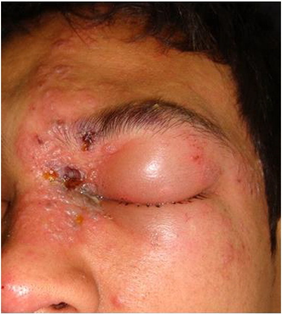
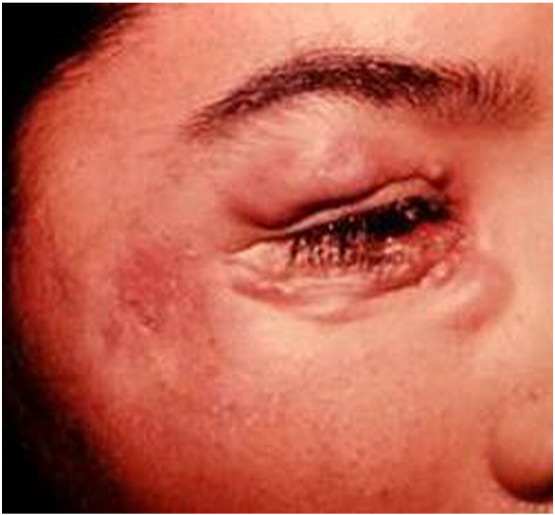
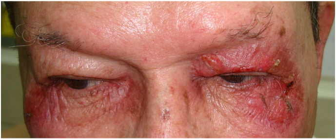
Herpes zoster.
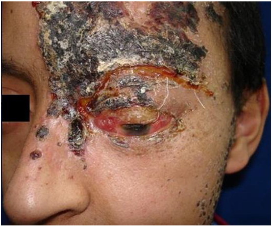
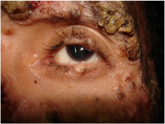
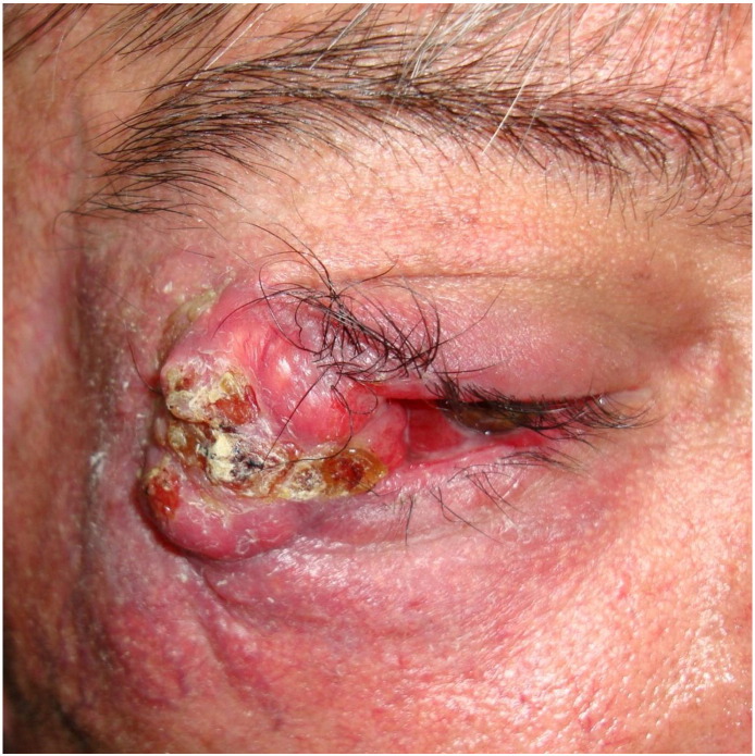
Severe necrosis and crusting originated by herpes zoster.
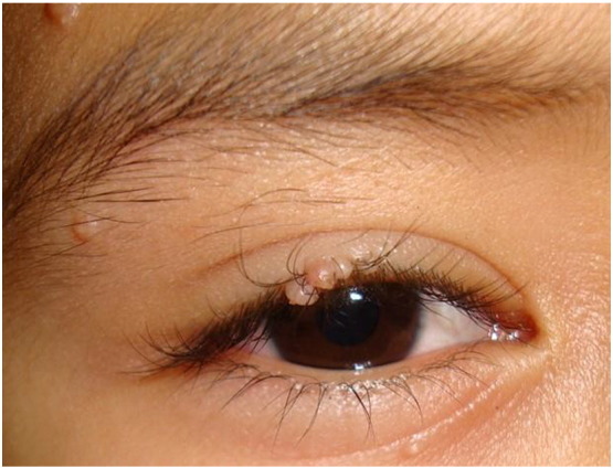
Molluscum contagiosum in the eyelash area.
Viral warts.
Sporotrichosis.
Histoplasmosis.
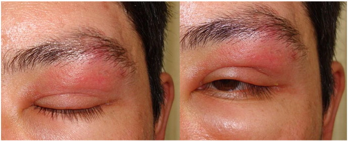
Urticarial and angioedema.
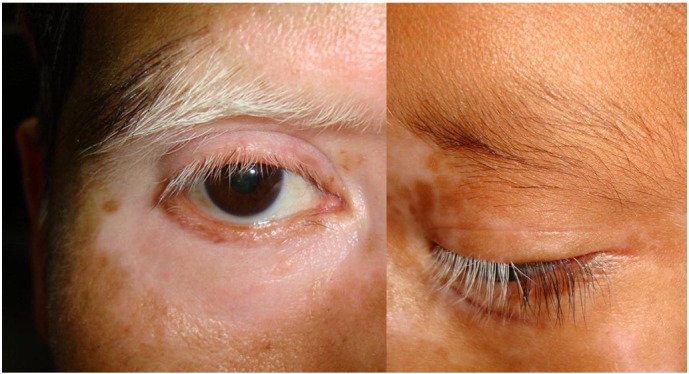
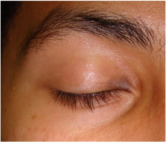
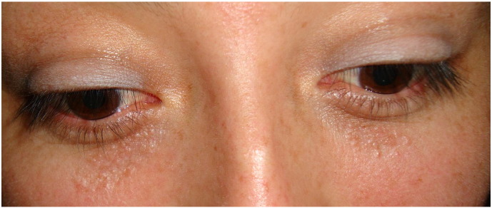
Vitiligo.
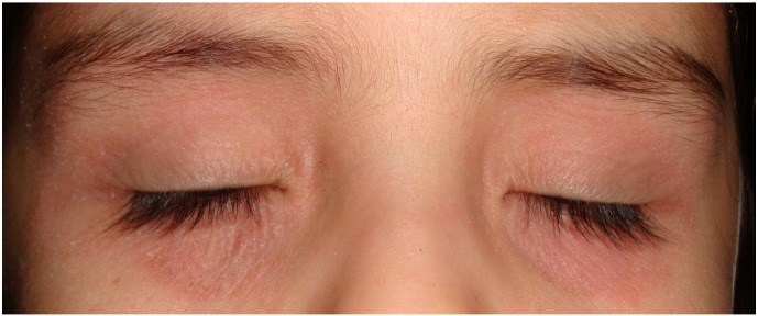
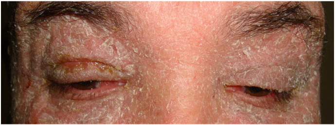
Atopic dermatitis.
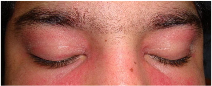
Dermatomyositis with the periocular rash (heliotrope rash) characteristic.
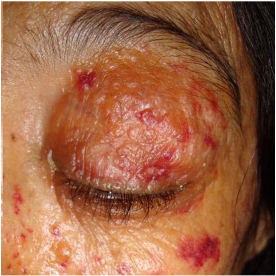
Amyloidosis with petechiae.
Fixed drug eruption to acetaminophen.
Stevens Johnson syndrome to albendazole.
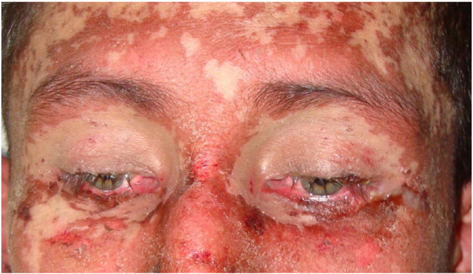
Stevens Johnson syndrome to allopurinol.
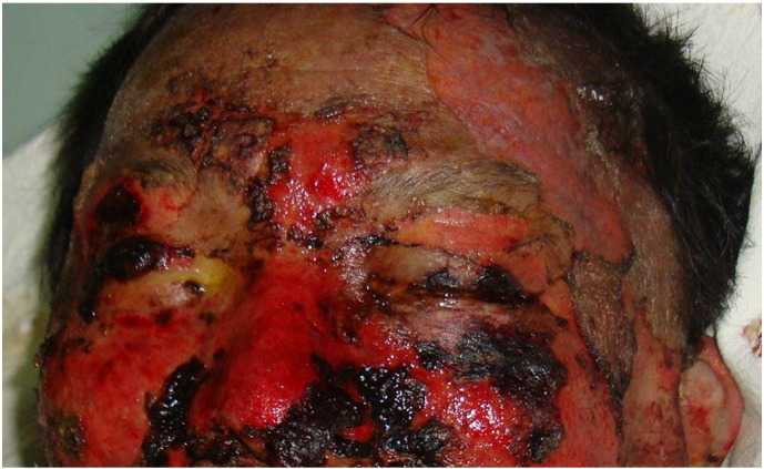
Toxic epidermal necrolysis to nonsteroidal anti-inflammatory drugs.
Erythroderma.
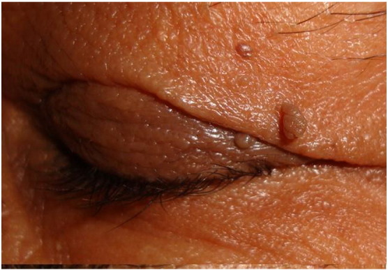
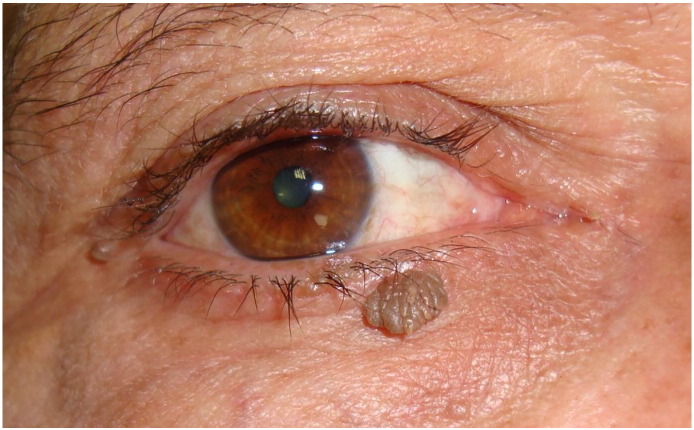
Acrochordons.
Syringomas.
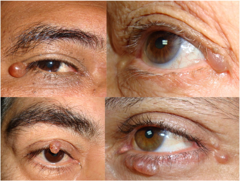
Hidrocystomas.
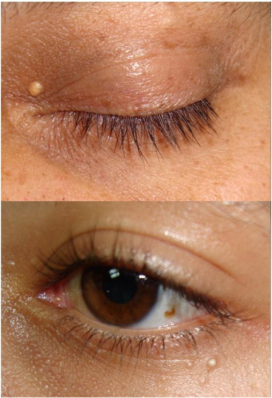
Milia cysts.
Seborrheic keratosis.
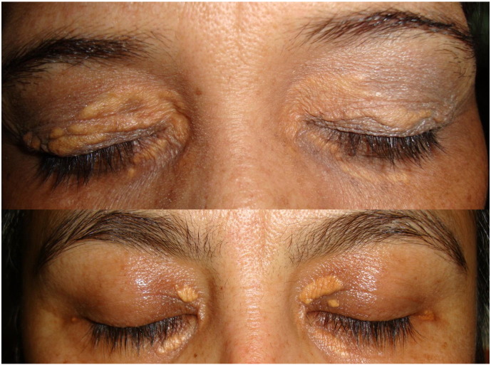
Xanthelasma.
Basal cell carcinoma.
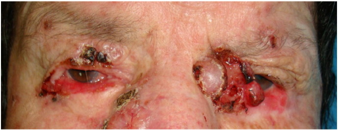
Squamous cell carcinoma.
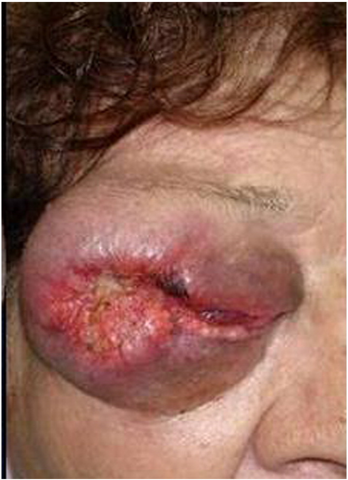
Sebaceous gland carcinoma.
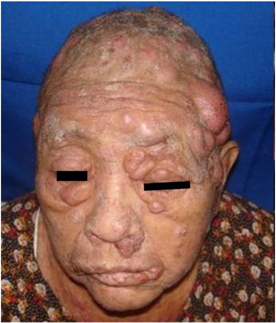
Mycosis fungoides.
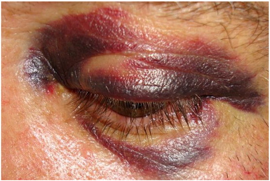
Hematoma.
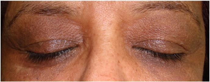
Dark circles under the eye.
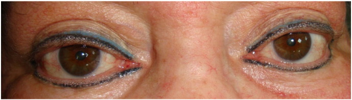
Cosmetic tattoo.
References
-
- Abalo-Lojo J.M., Cameselle-Teijeiro J., González F. Pilomatrixoma: Late onset in two periocular cases. Ophthal Plast Reconstr Surg. 2008;24:60–62. - PubMed
Publication types
LinkOut - more resources
Full Text Sources
Other Literature Sources
