

Vulvar lichen planus pemphigoides
- PMID: 29234717
- PMCID: PMC5715214
- DOI: 10.1016/j.ijwd.2017.07.002
Vulvar lichen planus pemphigoides
Abstract
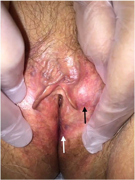
Lichen planus pemphigoides (LPP) is a rare blistering disease with features of both lichen planus and bullous pemphigoid. LPP typically appears on the extremities and occasionally involves the oral mucosa. Herein, we describe a case of LPP of the vulva of an 80-year-old woman, an uncommon location for this disease process. This clinical scenario can be confused with a number of similarly appearing entities such as erosive vulvar lichen planus, mucous membrane pemphigoid, and erosive lichen sclerosus et atrophicus. In fact, our patient carried a diagnosis of lichen sclerosus by an outside physician for 2 years prior to being properly diagnosed and treated. A detailed discussion of the epidemiology, clinical, and pathogenesis as well as the histologic and immunofluorescence characteristics of this uncommon diagnosis is presented. Our case emphasizes the necessity of microscopic analysis to differentiate lookalike disease states when making a diagnosis and choosing the correct therapeutics.
Figures


References
-
- Allen C.M., Camisa C., Grimwood R. Lichen planus pemphigoides: Report of a case with oral lesions. Oral Surg Oral Med Oral Pathol. 1987;63:184–188. - PubMed
-
- Archer C.B., Cronin E., Smith N.P. Diagnosis of lichen planus pemphigoides in the absence of bullae on normal-appearing skin. Clin Exp Dermatol. 1992;17:433–436. - PubMed
-
- Chan L.S., Ahmed A.R., Anhalt G.J., Bernauer W., Cooper K.D., Elder M.J. The first international consensus of mucous membrane pemphigoid. Arch Dermatol. 2002;138:370–379. - PubMed
-
- Cooper S.M., Dean D., Allen J., Kirtschig G., Wojnarowska F. Erosive lichen planus of the vulva: Weak circulating basement membrane zone antibodies are present. Clin Exp Dermatol. 2005;30:551–556. - PubMed
-
- Keith P.J., Wolz M.M., Peters M.S. Eosinophils in lichen sclerosus et atrophicus. J Cutan Pathol. 2015;42:693–698. - PubMed
Publication types
LinkOut - more resources
Full Text Sources
Other Literature Sources
