Review
. 2018 Apr;10(2):559-570.
doi: 10.1007/s12551-017-0378-z. Epub 2017 Dec 12.

Bacterial flagellar axial structure and its construction

Affiliations
Review

Bacterial flagellar axial structure and its construction

Katsumi Imada. Biophys Rev. 2018 Apr.

Abstract

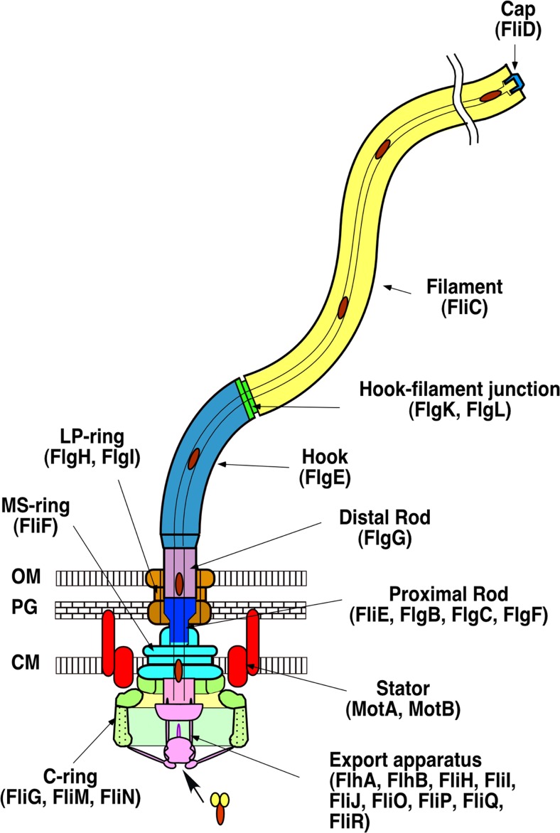
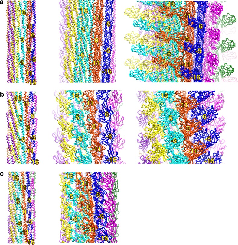
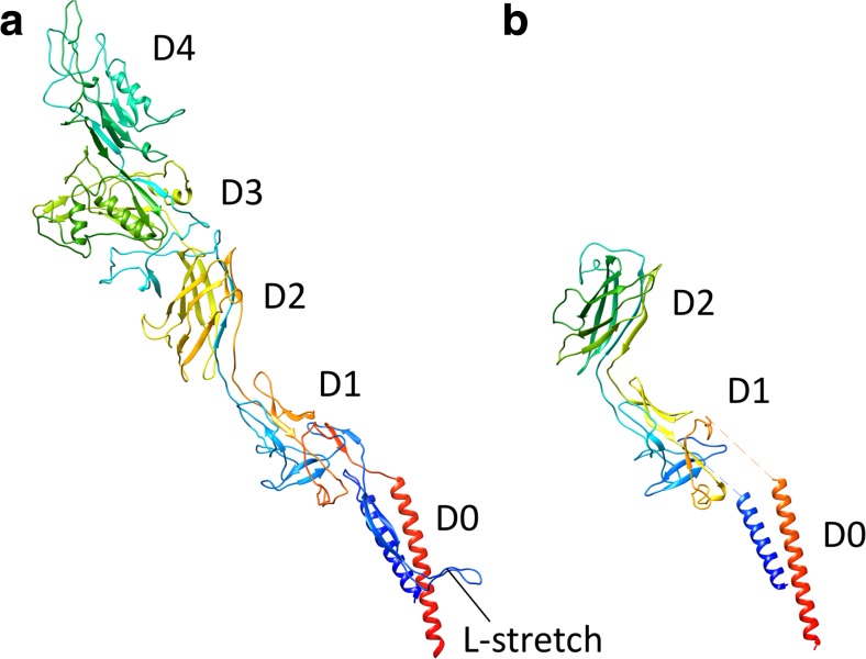
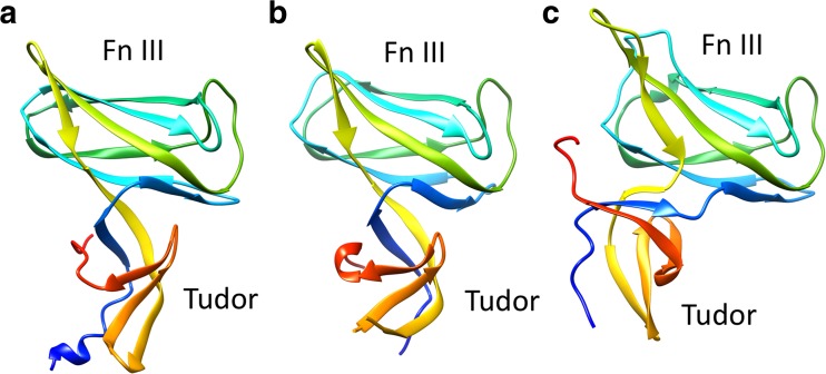
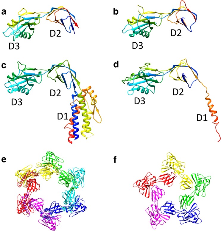
The bacterial flagellum is a motile organelle composed of thousands of protein subunits. The filamentous part that extends from the cell membrane is called the axial structure and consists of three major parts, the filament, hook, and rod, and other minor components. Each of the three main parts shares a similar self-assembly mechanism and a common basic architecture of subunit arrangement while showing quite distinct mechanical properties to achieve its specific function. Structural and molecular mechanisms to produce these various mechanical properties of the axial structure, such as the filament, the hook, and the rod, have been revealed by the complementary use of X-ray crystallography and cryo-electron microscopy. In addition, the mechanism of growth of the axial structure is beginning to be revealed based on the molecular structure.

Keywords: Axial structure; Bacterial flagellum; Cryo-electron microscopy; X-ray crystallography.


Conflict of interest statement

Conflict of interest

Katsumi Imada declares that he has no conflicts of interest.

Ethical approval

This article does not contain any studies with human participants or animals performed by the author.

Figures

Fig. 1
Fig. 1
Schematic drawing of the bacterial flagellum. Different colors represent different functional units. The names of the component proteins in each functional unit are shown in parentheses. CM Inner membrane, PG peptidoglycan layer, OM outer membrane
Fig. 2
Fig. 2
Common structural feature of the flagellar axial structure. a Arrangement of the protein subunits in the flagellar filament. Major helical lines are indicated by arrows. The subunits along the 11-start helical line comprise the protofilament. The number labeled on the subunits represents the number of the subunit starting from the central subunit (subunit 0) along the 1-start helical line. The number also shows the direction of the helical line. The subunit monomers are sequentially assembled along the 1-start helix. b Cross section of the flagellar filament structure. Each flagellin subunit is colored in the rainbow sequence of colors from blue to red for the – to C-terminus. The N- (blue) and C-terminal (red) regions form the inner tube. c Disordered regions of flagellar axial proteins. Disordered region and folded domains are colored in yellow and blue, respectively. The disordered regions were determined by limited proteolysis (Saijo-Hamano et al. 2004)
Fig. 3
Fig. 3
Structural comparison of flagellin from various bacteria. a Cryo-electron microscopy (Cryo-EM) structure of full-length flagellar filament protein, flagellin (FliC), from Salmonella (PDB ID 1ucu). bd Crystal structures of Burkholderia pseudomallei FliC (BpFliC; PDB ID 4cfi) (b), Pseudomonas aeruginosa FliC (PaFliC; PDB ID 4nx9) (c), and Bacillus subtilis FliC (BsFliC; (PDB ID 5gy2) (d). The D0 domains of Bp-, Pa and Bs-FliC were removed for crystallization. The chains are colored in the rainbow sequence of colors from blue to red for the N- to C-terminus
Fig. 4
Fig. 4
Comparison of the protofilament structures of the filament (a), the hook (b), and the rod (c). Two protein subunits in a protofilament are shown in each panel. The chains are colored in the rainbow sequence of colors from blue to red for the N- to C-terminus. The D0 and D1 helices of FliC are arranged nearly parallel to the filament axis and densely packed along the protofilament. The D0 helices of flagellar hook protein FlgE are tilted to form a gap between the axially neighboring subunits (blue two-direction arrow). The N-terminal helix of flagellar rod protein FlgG is one turn longer than that of FlgE; therefore the gap between the N-terminal helix and the axially neighboring subunit (red two-direction arrow) is shorter than that in the hook (green two-direction arrow). The D1 domains of FlgG are arranged upright to make an interaction with the neighboring subunit (magenta arrow), while those of FlgE in the hook are tilted, producing an axial gap along the protofilament (black two-direction arrow)
Fig. 5
Fig. 5
Intersubunit domain interactions in the axial structure. Side views of the R-type filament (a), the hook (b), and the rod (c) showing the subunit arrangement and packing of the different layers: left panel shows the array of the D0 domains; middle panel shows the D1 domains; right panel shows the D2 and D3 domains. Individual protofilaments are shown in different colors. The subunits surrounding subunit 0 are labeled with the number showing the direction of the helical line
Fig. 6
Fig. 6
Structural comparison of the Campylobacter jejuni flagellar hook protein FlgE (CjFlgE; PDB ID 5jxl) (a) and Salmonella enterica serovar Typhimurium FlgE (StFlgE; PDB ID 3A69) (b). The chains connecting the D0 and D1 domains are not determined in the StFlgE structure. The chains are drawn in rainbow sequence of colors from blue to red for the N- to C-terminus
Fig. 7
Fig. 7
Crystal structures of the C-terminal domain of FlgD (FlgDc), a scaffolding protein for flagellar hook assembly, from various bacteria. a Xanthomonas campestris (XcFlgDc; PDB ID 3c12), b Pseudomonas aeruginosa (PaFlgDc; PDB ID 3osv), c Helicobacter pylori (HpFlgDc; PDB ID 4zzf). The chains are shown in rainbow sequence of colors from blue to red for the N- to C-terminus
Fig. 8
Fig. 8
Crystal structures of the capping protein of the bacterial flagellar filament (FliD) from various bacteria. Ribbon drawings of StFliD (PDB ID 5h5t) (a), Serratia marcescens FliD (SmFliD; PDB ID 5xlk) (b), Escherichia coli FliD (EcFliD; PDB ID 5h5v) (b), and PaFliD (PDB ID 5fhy) (d) are shown in rainbow sequence of colors from blue to red for the N- to C-terminus. e, f Structure of the FliD cap assembly: e EcFliD hexamer (PDB ID 5h5v), f StFliD pentamer (PDB ID 5h5t). Subunits are represented in different colors

References

    1. Asakura S. Polymerization of flagellin and polymorphism of flagella. Advan Biophys (Japan) 1970;1:99–155. - PubMed
    1. Berg HC, Anderson RA. Bacteria swim by rotating their flagellar filaments. Nature. 1973;245:380–382. doi: 10.1038/245380a0. - DOI - PubMed
    1. Blair DF, Berg HC. Restoration of torque in defective flagellar motors. Science. 1988;242:1678–1681. doi: 10.1126/science.2849208. - DOI - PubMed
    1. Buist G, Kok J, Leenhouts KJ, Dabrowska M, Venema G, Haandrikman AJ. Molecular cloning and nucleotide sequence of the gene encoding the major peptidoglycan hydrolase of Lactococcus lactis, a muramidase needed for cell separation. J Bacteriol. 1995;177:1554–1563. doi: 10.1128/jb.177.6.1554-1563.1995. - DOI - PMC - PubMed
    1. Calladine CR. Construction of bacterial flagella. Nature. 1975;225:121–124. doi: 10.1038/255121a0. - DOI - PubMed

LinkOut - more resources