. 2018 Jun;16(2):253-261.
doi: 10.1111/vco.12375. Epub 2017 Dec 13.

Anticancer effects of resveratrol in canine hemangiosarcoma cell lines

Affiliations

Anticancer effects of resveratrol in canine hemangiosarcoma cell lines

A Carlson et al. Vet Comp Oncol. 2018 Jun.

Erratum in

  • Corrigendum.
    [No authors listed] [No authors listed] Vet Comp Oncol. 2018 Dec;16(4):677. doi: 10.1111/vco.12445. Epub 2018 Oct 4. Vet Comp Oncol. 2018. PMID: 30421502 No abstract available.

Abstract

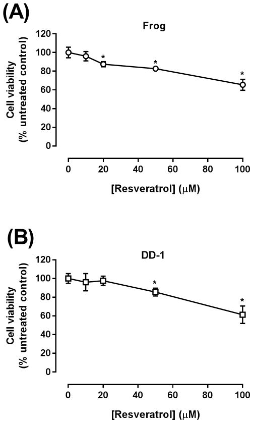
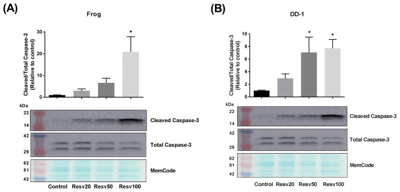
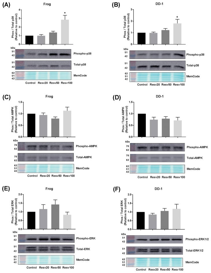
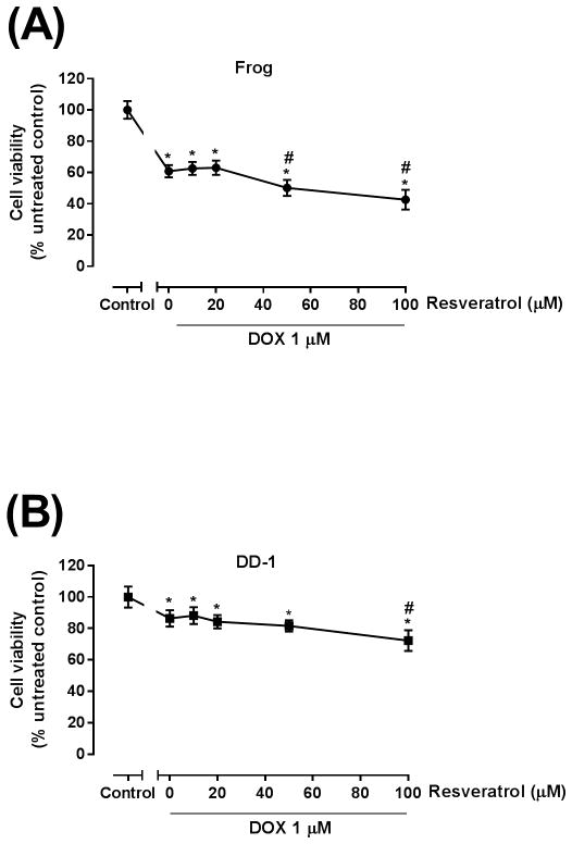
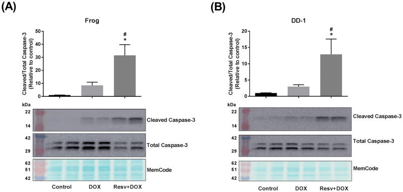
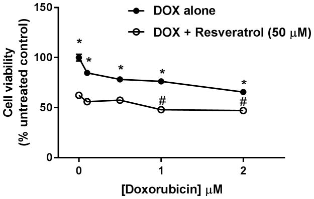
Hemangiosarcoma (HSA) is a highly malignant tumour with aggressive biological behaviour. HSAs are more common in dogs than other domestic animals. The median survival time of dogs with HSA remains short, even with chemotherapy and surgery. Therefore, there is a critical need to improve the adjuvant chemotherapeutic regimens to improve clinical outcomes in dogs with HSA. Resveratrol has been shown to possess strong anti-proliferative and/or pro-apoptotic properties in human cancer cell lines. Nevertheless, the potential anticancer effects of resveratrol have not been reported in canine HSAs. The objective of this study is to determine the growth inhibitory effects of resveratrol in HSA cells when used alone or in combination with doxorubicin, a commonly used chemotherapeutic agent. Frog and DD-1 canine HSA cell lines were treated with varying concentrations of resveratrol with and without doxorubicin. Cell viability was measured by the MTT assay. The expression of apoptotic proteins, activation of p38 mitogen-activated protein kinase (MAPK), AMP-activated protein kinase (AMPK) and extracellular signal-regulated kinase 1/2 (ERK1/2) were assessed by western blotting. Similar to human cancer cell lines, resveratrol markedly inhibited the growth and induced apoptosis in both HSA cell lines. Mechanistically, resveratrol activated p38 MAPK, but did not affect the AMPK or the ERK1/2 pathways. Additional experiments showed that resveratrol augmented the growth-inhibitory and apoptotic effects of doxorubicin in both HSA cell lines. These findings suggest that resveratrol has pro-apoptotic effects in canine HSA cells; therefore, its use as a potential adjunct therapy in canine HSA patients warrants further investigation.

Keywords: apoptosis; canine; doxorubicin; hemangiosarcoma; resveratrol.


Figures

Figure 1
Figure 1. Resveratrol inhibits the growth of Frog (A) and DD-1 cells (B)
Canine HSA cells were incubated with increasing concentrations of resveratrol (10–100 μM) for 24 h in Frog cells and for 48 h in DD-1 cells. Cell viability was determined by the MTT assay. Results are shown as Mean +/− SEM (n = 8). * p < 0.05 as compared to control values by one-way ANOVA.
Figure 2
Figure 2. Resveratrol (Resv) induces apoptosis in Frog (A) and DD-1 cells (B)
Canine HSA cells were incubated with increasing concentrations of resveratrol (20, 50, and 100 μM) for 24 h. Induction of apoptosis was assessed by measuring the cleaved caspase-3/total caspase-3 by western blotting. Results are shown as Mean +/− SEM (n = 6) from three independent experiments (duplicate of wells on three independent times). * p < 0.05 as compared to control values by one-way ANOVA.
Figure 3
Figure 3. Resveratrol (Resv) activates p38 MAPK (A and B), but does not affect AMPK (C and D) or ERK1/2 (E and F) pathways
Canine HSA cells were incubated with increasing concentrations of resveratrol (20, 50, and 100 μM) for 24 h. p38 MAPK activation was assessed by measuring the phospho p38 MAPK/total p38 MAPK in Frog (A) or DD-1 (B). AMPK activation was assessed by measuring the phospho-AMPK/total AMPK in Frog (C) or DD-1 (D) cells. Likewise, ERK1/2 activation was assessed by measuring the phospho-ERK1/2/total ERK1/2 in Frog (E) or DD-1 (F) cells. Results are shown as Mean +/− SEM (n = 6) from three independent experiments (duplicate of wells on three independent times). * p < 0.05 as compared to control values by one-way ANOVA.
Figure 4
Figure 4. Resveratrol increased the growth inhibitory effect of DOX in Frog (A) and DD-1 cells (B)
Canine HSA cells were incubated with 1 μM DOX in the absence or presence of increasing concentrations of resveratrol (10–100 μM) for 24 h in Frog cells and for 48 h in DD-1 cells. Cell viability was determined by the MTT assay. Results are shown as Mean +/− SEM (n = 8). * p < 0.05 as compared to control, # p < 0.05 as compared to doxorubicin-treated cells by one-way ANOVA.
Figure 5
Figure 5. Resveratrol (Resv) augments doxorubicin (DOX)-induced apoptosis in Frog (A) and DD-1 cells (B)
Canine HSA cells were incubated with 100 μM resveratrol for 2 h, then treated with 1 μM DOX for an additional 22 h. Induction of apoptosis was assessed by measuring the cleaved caspase-3/total caspase-3 by western blotting. Results are shown as Mean +/− SEM (n = 6) from three independent experiments (duplicate of wells on three independent times). * p < 0.05 as compared to control values, # p < 0.05 as compared to cells treated by doxorubicin only by one-way ANOVA.
Figure 6
Figure 6. Resveratrol increased the growth inhibitory effect of a broad range of doxorubicin (DOX) concentrations in Frog cells
Canine HSA Frog cells were incubated with 0.1 – 2 μM DOX in the absence or presence of 50 μM resveratrol for 24 h. Cell viability was determined by the MTT assay. Results are shown as Mean +/− SEM (n = 8). * p < 0.05 as compared to resveratrol-treated cells, # p < 0.05 as compared to cells treated by resveratrol only by one-way ANOVA.
Figure 7
Figure 7. Resveratrol does not increase doxorubicin (DOX) toxicity in H9c2 cardiomyoblast cell line
H9c2 cells were incubated with 1 μM DOX in the absence or presence of increasing concentrations of resveratrol (10–100 μM) for 24 h. Cell viability was determined by the MTT assay. Results are shown as Mean +/− SEM (n = 8). * p < 0.05 as compared to control by one-way ANOVA.

References

    1. Lamerato-Kozicki AR, Helm KM, Jubala CM, Cutter GC, Modiano JF. Canine hemangiosarcoma originates from hematopoietic precursors with potential for endothelial differentiation. Exp Hematol. 2006;34:870–8. - PubMed
    1. Finotello R, Stefanello D, Zini E, Marconato L. Comparison of doxorubicin-cyclophosphamide with doxorubicin-dacarbazine for the adjuvant treatment of canine hemangiosarcoma. Vet Comp Oncol. 2017;15:25–35. - PubMed
    1. Goritz M, Muller K, Krastel D, et al. Canine splenic haemangiosarcoma: influence of metastases, chemotherapy and growth pattern on post-splenectomy survival and expression of angiogenic factors. J Comp Pathol. 2013;149:30–9. - PubMed
    1. Wendelburg KM, Price LL, Burgess KE, et al. Survival time of dogs with splenic hemangiosarcoma treated by splenectomy with or without adjuvant chemotherapy: 208 cases (2001–2012) J Am Vet Med Assoc. 2015;247:393–403. - PubMed
    1. Wood CA, Moore AS, Gliatto JM, et al. Prognosis for dogs with stage I or II splenic hemangiosarcoma treated by splenectomy alone: 32 cases (1991–1993) J Am Anim Hosp Assoc. 1998;34:417–21. - PubMed