. 2017 Dec 13;7(1):17525.
doi: 10.1038/s41598-017-17758-4.

In vivo metabolic imaging of Traumatic Brain Injury

Affiliations

In vivo metabolic imaging of Traumatic Brain Injury

Caroline Guglielmetti et al. Sci Rep. .

Abstract

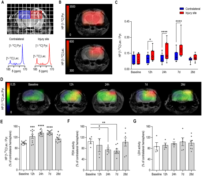
Complex alterations in cerebral energetic metabolism arise after traumatic brain injury (TBI). To date, methods allowing for metabolic evaluation are highly invasive, limiting our understanding of metabolic impairments associated with TBI pathogenesis. We investigated whether 13C MRSI of hyperpolarized (HP) [1-13C] pyruvate, a non-invasive metabolic imaging method, could detect metabolic changes in controlled cortical injury (CCI) mice (n = 57). Our results show that HP [1-13C] lactate-to-pyruvate ratios were increased in the injured cortex at acute (12/24 hours) and sub-acute (7 days) time points after injury, in line with decreased pyruvate dehydrogenase (PDH) activity, suggesting impairment of the oxidative phosphorylation pathway. We then used the colony-stimulating factor-1 receptor inhibitor PLX5622 to deplete brain resident microglia prior to and after CCI, in order to confirm that modulations of HP [1-13C] lactate-to-pyruvate ratios were linked to microglial activation. Despite CCI, the HP [1-13C] lactate-to-pyruvate ratio at the injury cortex of microglia-depleted animals at 7 days post-injury remained unchanged compared to contralateral hemisphere, and PDH activity was not affected. Altogether, our results demonstrate that HP [1-13C] pyruvate has great potential for in vivo non-invasive detection of cerebral metabolism post-TBI, providing a new tool to monitor the effect of therapies targeting microglia/macrophages activation after TBI.


Conflict of interest statement

The authors declare that they have no competing interests.

Figures

Figure 1
Figure 1
Longitudinal evaluation of TBI using high field T2-weighted MR imaging. (A) Representative T2-weigthed MR images were acquired prior to injury (Baseline), twelve hours (12 h), twenty-four hours (24 h), seven days (7 d) and twenty-eight days (28 d) post-injury. Cortical alterations of the tissue microstructure can be detected as a mix of hypo-intense and hyper-intense contrast in the injured hemisphere as early as 12 h post-injury, and persist until 28 d post-injury. The formation of a well-defined cavity can be observed at 7 d/28 d post-injury. (B) Representative 3D reconstruction of the lesioned area (red), cavitation (blue) and ventricles (yellow) for each time point. (C) Quantitative analyses revealed highest lesion size at acute time points following injury (12 h and 24 h, p < 0.0001, compared to Baseline). The lesioned area decreased by 53 ± 9% at 7 d (p = 0.0003, compared to 24 h) and persists until 28 d post-injury (p = 0.0005, compared to Baseline). The formation of a cavitation can be observed by 28 days post-injury (p = 0.0174, compared to Baseline). All values are reported as mean ± sem (n = 5 mice).
Figure 2
Figure 2
Longitudinal MR metabolic imaging following TBI reveals a transient increase of the HP [1-13C] lactate-to-pyruvate ratio, which is paralleled by decreased PDH activity. (A) Representative T2-weighted image with overlaid grid used for the acquisition of HP 13C MRSI, highlighting the voxels containing the injured (red) and contralateral (blue) hemispheres. HP 13C spectra for the contralateral hemisphere (blue) and injured hemisphere (red) showed increased HP [1-13C] lactate in the injured hemisphere at 7 days post-injury. (B) Corresponding heatmaps of HP [1-13C] pyruvate and HP [1-13C] lactate showed even distribution of HP [1-13C] pyruvate within the brain while HP [1-13C] lactate is increased at the level of the injury. (C) Quantitative analyses of the HP [1-13C] lactate-to-pyruvate ratios revealed a significant increase at 12 h, 24 h and 7 d post-injury between injured and contralateral hemispheres (Two-Way ANOVA, p < 0.0001 for hemisphere effect, p = 0.0259 for time effect, p = 0.0002 for hemisphere and time interaction). (D) Representative heatmaps indicating the highest level of the HP [1-13C] lactate-to-pyruvate ratio at the site of injury. (E) HP [1-13C] lactate-to-pyruvate ratio expressed as a percent change of the contralateral hemisphere showed increased ratios by 23 ± 7% at 12 hours (p = 0.0002), 36 ± 3% at 24 hours (p < 0.0001), 37 ± 2% at 7 days (p < 0.0001) and 15 ± 4% at 28 days post injury (p = 0.0132) compared to Baseline. (F) PDH activity was significantly decreased in the injured hemisphere by 30 ± 6% at 24 h (p = 0.0358) and 35 ± 7% at 7 d post-injury (p = 0.0011) while LDH activity remained unchanged (G). All values are reported as mean ± sem (n = 10 mice for HP [1-13C] MRSI, n = 4–8 mice per time point for PDH and LDH activity).
Figure 3
Figure 3
Immunofluorescence analyses following TBI and PLX5622 treatment. (A) Representative immunofluorescence images taken at 7 d post-injury showing a strong reduction of microglia/macrophages (Iba-1, red; CD11b, green), and CD68 expressing microglia/macrophages (yellow) in the group that received the PLX5622 compared to control diet. Quantitative analyses in the injured cortex confirmed decrease of (B) Iba-1 expressing microglia/macrophage population (92 ± 7%, p < 0.0001), (C) CD11b expressing microglia/macrophages (83 ± 5%, p = 0.0001), (D) the lysosomal marker CD68 (76 ± 4%, p = 0.0014), in the group that received the PLX5622 diet. Similarly, quantitative analyses in the contralateral cortex showed decrease of (E) Iba-1 and (F) CD11b expressing microglia/macrophage population (84 ± 7%, p < 0.0082 and 87 ± 6%, p < 0.0019, respectively) for mice that received PLX5622 compared to control diet, while the (G) lysosomal marker CD68 levels were below detection. All values are reported as mean ± sem (n = 3–5 mice per group).
Figure 4
Figure 4
The HP [1-13C] lactate-to-pyruvate ratio is sensitive to modulation of microglia/macrophages status following TBI. (A) Representative heatmaps of the HP [1-13C] lactate-to-pyruvate ratios and corresponding HP 13C spectra showing increased HP [1-13C] lactate-to-pyruvate ratio and increased HP [1-13C] lactate production at 7d post-injury in the mouse that received the control diet but not in the mouse that received the PLX5622 diet. (B) Quantitative analyses of the HP [1-13C] lactate-to-pyruvate ratios revealed no significant differences between the injured and contralateral hemispheres of microglia depleted mice (Two-Way ANOVA, p = 0.089 for hemisphere effect, p = 0.1804 for time effect, p = 0.2792 for hemisphere and time interaction) at 7 days post-injury, contrasting with the increase of HP [1-13C] lactate-to-pyruvate ratio observed in mice that received the control diet (p = 0.0001). (C) HP [1-13C] lactate-to-pyruvate ratio, expressed as a percent change of the contralateral hemisphere, showed no change of the HP [1-13C] lactate-to-pyruvate ratio at 7 d post-injury compared to Baseline in mice that received the PLX5622, in contrast with the 37 ± 2% at 7 d (p < 0.0001) in mice that received the control diet. (D) PDH activity was decreased in the injured hemisphere of mice that received a control diet (p = 0.0011) but not in mice that received PLX5622 diet (p = 0.9429). (E) LDH activity remained unchanged (p = 0.1769). All values are reported as mean ± sem (n = 5–10 mice per group).

References

    1. Kumar A, Loane DJ. Neuroinflammation after traumatic brain injury: opportunities for therapeutic intervention. Brain Behav Immun. 2012;26:1191–1201. doi: 10.1016/j.bbi.2012.06.008. - DOI - PubMed
    1. Masel BE, DeWitt DS. Traumatic brain injury: a disease process, not an event. J Neurotrauma. 2010;27:1529–1540. doi: 10.1089/neu.2010.1358. - DOI - PubMed
    1. Lozano D, et al. Neuroinflammatory responses to traumatic brain injury: etiology, clinical consequences, and therapeutic opportunities. Neuropsychiatr Dis Treat. 2015;11:97–106. - PMC - PubMed
    1. Simon DW, et al. The far-reaching scope of neuroinflammation after traumatic brain injury. Nat Rev Neurol. 2017;13:171–191. doi: 10.1038/nrneurol.2017.13. - DOI - PMC - PubMed
    1. Morganti JM, et al. Age exacerbates the CCR2/5-mediated neuroinflammatory response to traumatic brain injury. J Neuroinflammation. 2016;13:80. doi: 10.1186/s12974-016-0547-1. - DOI - PMC - PubMed

Publication types

MeSH terms