. 2017 Dec 13;7(1):17489.
doi: 10.1038/s41598-017-17792-2.

Biodegradable Bisvinyl Sulfonemethyl-crosslinked Gelatin Conduit Promotes Regeneration after Peripheral Nerve Injury in Adult Rats

Affiliations

Biodegradable Bisvinyl Sulfonemethyl-crosslinked Gelatin Conduit Promotes Regeneration after Peripheral Nerve Injury in Adult Rats

Chien-Hsin Ko et al. Sci Rep. .

Abstract

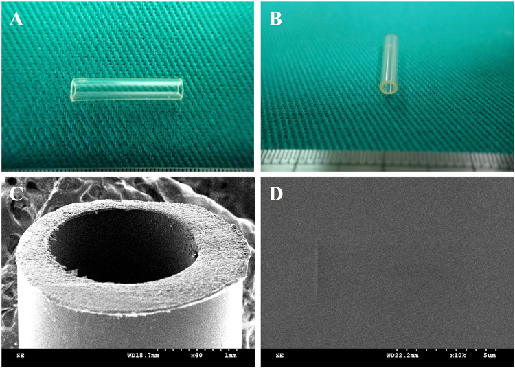
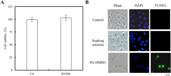
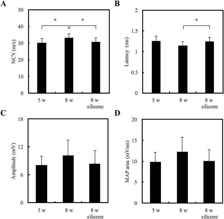
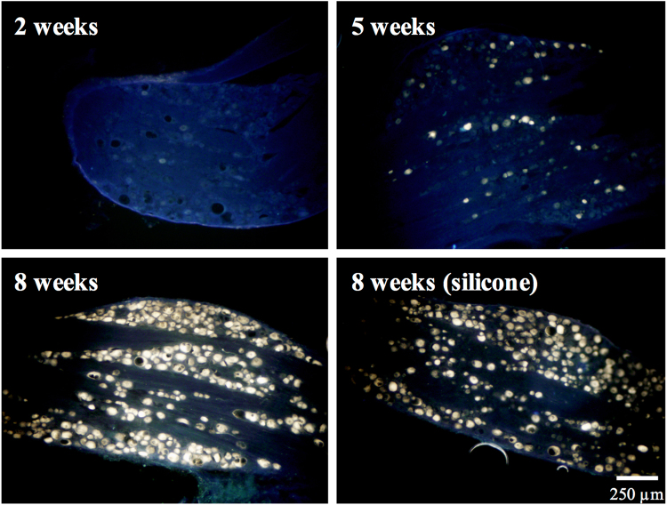
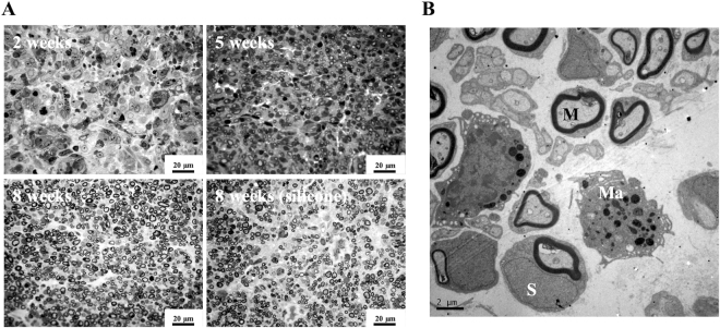
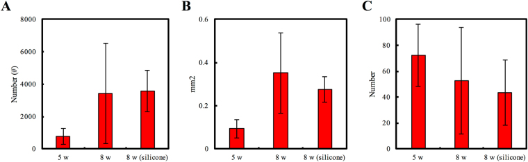
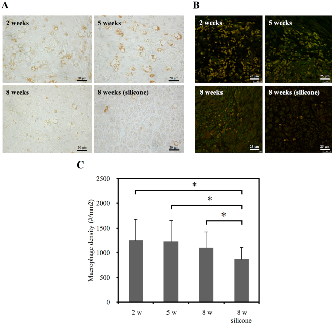
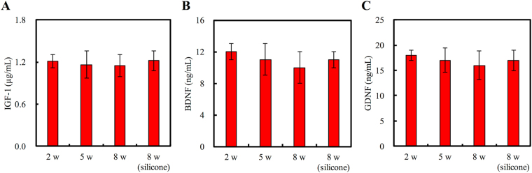
In our previous study, we found that gelatin-based materials exhibit good conductivity and are non-cytotoxic. In this study, gelatin was cross-linked with bisvinyl sulfonemethyl (BVSM) to fabricate a biodegradable conduit for peripheral nerve repair. First, BVSM on the prepared conduit was characterized to determine its mechanical properties and contact angle. The maximum tensile strength and water contact angle of the gelatin-BVSM conduits were 23 ± 4.8 MPa and 74.7 ± 9°, which provided sufficient mechanical strength to resist muscular contraction; additionally, the surface was hydrophilic. Cytotoxicity and apoptosis assays using Schwann cells demonstrated that the gelatin-BVSM conduits are non-cytotoxic. Next, we examined the neuronal electrophysiology, animal behavior, neuronal connectivity, macrophage infiltration, calcitonin gene-related peptide localization and expression, as well as the expression levels of nerve regeneration-related proteins. The number of fluorogold-labelled cells and histological analysis of the gelatin-BVSM nerve conduits was similar to that observed with the clinical use of silicone rubber conduits after 8 weeks of repair. Therefore, our results demonstrate that gelatin-BVSM conduits are promising substrates for application as bioengineered grafts for nerve tissue regeneration.


Conflict of interest statement

The authors declare that they have no competing interests.

Figures

Figure 1
Figure 1
(A,B) Overview, optical micrographs, and SEM micrograph with (C) low and (D) high magnification of BVSM-gelatin conduits.
Figure 2
Figure 2
Induction of biocompatibility using extract solution of BVSM-gelatin conduits. (A) Quantification of cytotoxic test of extract solutions of BVSM-gelatin conduits relative to controls on Schwann cells. (B) Nuclei of Schwann cells were characterized by DAPI and TUNEL assay and investigated under a fluorescence microscope. Scale bars: 50 µm.
Figure 3
Figure 3
NF-κB-dependent bioluminescence in living mice implanted with BVSM-gelatin conduits. (A) Diagrams show the bioluminescent signal within a radius of 2.5 mm of implanted region (boxed area). The color overlay on the image represents the photons s21 emitted from the animal, as indicated by the color scales. (B) Quantification of photon emission within the implanted region.
Figure 4
Figure 4
(A) Weight loss and (B) micrograph of interface area of BVSM-gelatin conduits after implantation for different times. Scale bars: 100 µm.
Figure 5
Figure 5
Analysis of evoked muscle action potentials, including (A) NCV, (B) latency, (C) peak amplitude, and (D) MAP area. *Indicates a significant difference (P < 0.05) from other examined time points.
Figure 6
Figure 6
Representative images of the retrograde axonal tracing with fluorogold for different time points. Scale bar: 250 μm.
Figure 7
Figure 7
(A) Representative micrographs of cross-sections of regenerated sciatic nerves. (B) Ultrastructural observation using TEM to show macrophage (Ma), myelinated axon (M), and Schwann cell (S) infiltration and remyelination in regenerated nerves.
Figure 8
Figure 8
Morphometric analysis of regenerated nerves in the BVSM-gelatin conduits after implantation for 5 and 8 weeks, (A) axon number, (B) total nerve area and (C) blood vessel number.
Figure 9
Figure 9
(A) Photomicrographs demonstrating anti-rat CD68 immunoreactivity in macrophages from cross-sections of distal nerve cables after BVSM-gelatin or silicone rubber conduits implanted for different times. (B) Representative photographs of CD68 and Iba-1 immunoreactivity on the macrophages. (C) Quantitation of macrophage infiltration density. *Indicates a significant difference (P < 0.05) from other examined time points.
Figure 10
Figure 10
(A) Representative histological micrographs of CGRP expression and (B) Quantitation of the ratio of CGRP expression area. *Indicates a significant difference (P < 0.05) from other examined time points.
Figure 11
Figure 11
Effects of BVSM-gelatin conduits on proteins expression of (A) IGF-1, (B) BDNF, and (C) GDNF. Values represent the means ± standard deviation (SD) for 10 rats in each group.

References

    1. Cheney FW, Domino K, Caplan RA, Posner KL. Nerve injury associated with anesthesia. Anesthesiology. 1990;90:1062–1069. doi: 10.1097/00000542-199904000-00020. - DOI - PubMed
    1. Kouyoumdjian JA. Peripheral nerve injuries: A retrospective survey of 456 cases. Muscle & Nerve. 2006;34:785–788. doi: 10.1002/mus.20624. - DOI - PubMed
    1. Tajdaran K, Gordon T, Wood MD, Shoichet MS, Borschel GH. A glial cell line-derived neurotrophic factor delivery system enhances nerve regeneration across acellular nerve allografts. Acta Biomater. 2016;29:62–70. doi: 10.1016/j.actbio.2015.10.001. - DOI - PubMed
    1. Johnson BN, et al. 3D printed anatomical nerve regeneration pathways. Adv Funct Mater. 2015;25:6205–6217. doi: 10.1002/adfm.201501760. - DOI - PMC - PubMed
    1. Longo MVL, et al. Comparisons of the results of peripheral nerve defect repair with fibrin conduit and autologous nerve graft: An experimental study in rats. Microsurgery. 2016;36:59–65. doi: 10.1002/micr.22413. - DOI - PubMed

Publication types

MeSH terms