Review
. 2018 Apr;19(4):229-244.
doi: 10.1038/nrm.2017.119. Epub 2017 Dec 13.

Ten principles of heterochromatin formation and function

Affiliations
Review

Ten principles of heterochromatin formation and function

Robin C Allshire et al. Nat Rev Mol Cell Biol. 2018 Apr.

Abstract

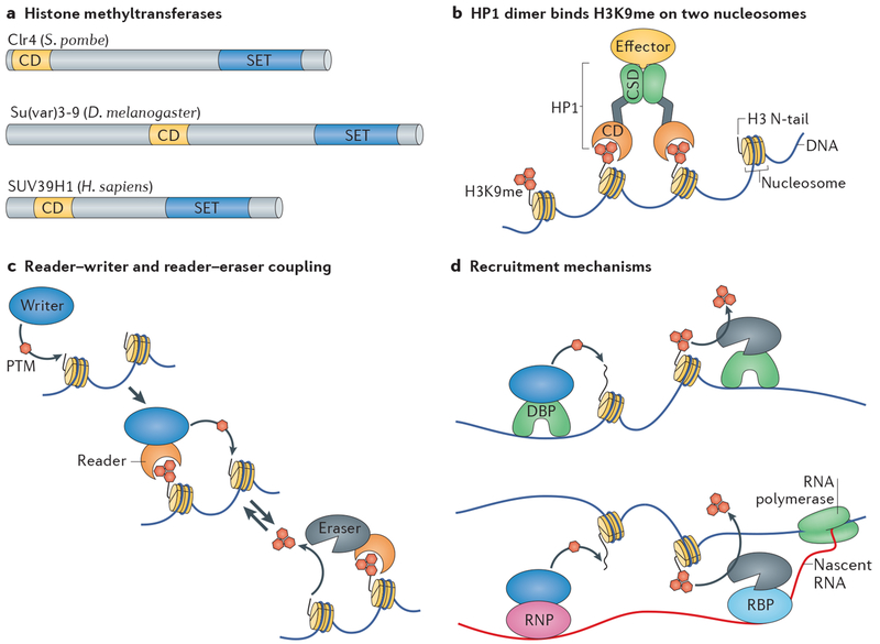
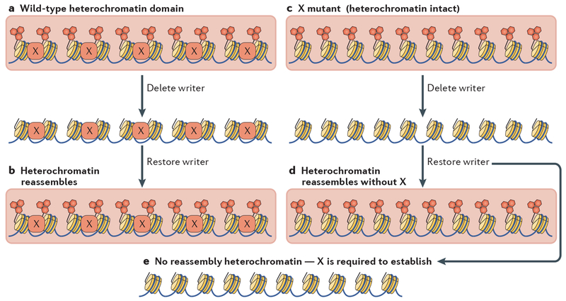
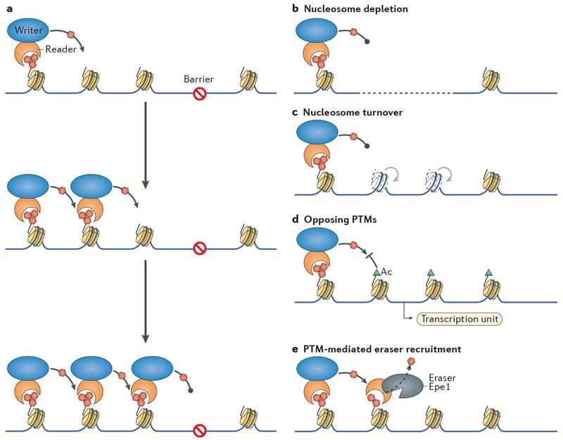
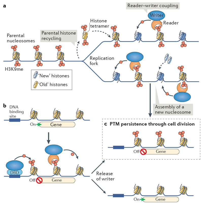
Heterochromatin is a key architectural feature of eukaryotic chromosomes, which endows particular genomic domains with specific functional properties. The capacity of heterochromatin to restrain the activity of mobile elements, isolate DNA repair in repetitive regions and ensure accurate chromosome segregation is crucial for maintaining genomic stability. Nucleosomes at heterochromatin regions display histone post-translational modifications that contribute to developmental regulation by restricting lineage-specific gene expression. The mechanisms of heterochromatin establishment and of heterochromatin maintenance are separable and involve the ability of sequence-specific factors bound to nascent transcripts to recruit chromatin-modifying enzymes. Heterochromatin can spread along the chromatin from nucleation sites. The propensity of heterochromatin to promote its own spreading and inheritance is counteracted by inhibitory factors. Because of its importance for chromosome function, heterochromatin has key roles in the pathogenesis of various human diseases. In this Review, we discuss conserved principles of heterochromatin formation and function using selected examples from studies of a range of eukaryotes, from yeast to human, with an emphasis on insights obtained from unicellular model organisms.


Conflict of interest statement

Competing interests statement

The authors declare no competing interests.

Figures

Figure 1 |
Figure 1 |. Core heterochromatin components and mechanisms.
a | The protein domain organization of the histone–lysine N-methyltransferases (KMTs) cryptic loci regulator 4 (Clr4) of Schizosaccharomyces pombe, suppressor of variegation 3-9 (Su(var)3-9) of Drosophila melanogaster and SUV39H1 (SU(VAR)3-9 homologue 1) of Homo sapiens. The SET (Su(var)3-9, enhancer of zeste and trithorax) domain isthe KMT catalytic domain and uses S-adenosyl-methionine as a methyl donor to methylate histone H3 lysine 9. The chromodomain (CD) specifically recognizes methylated histone H3 lysine 9 (H3K9me). b | Depiction of a heterochromatin protein 1 (HP1) dimer bound to nucleosomes modified with H3K9me (red hexagons). The chromodomain and the chromoshadow domain (CSD), which is a dimerization domain, of HP1 are shown. The platform produced by the CSD dimer enables binding of effector proteins. For simplicity, only one of the two H3 tails that protrude from the octamer core is shown on each nucleosome. c | Heterochromatin assembly and disassembly by reader-modifier coupling. Different ‘writer’ enzymes catalyse the addition of a post-translational modification (PTM) to a histone within a nucleosome, whereas ‘eraser’ enzymes catalyse the removal of PTMs. ‘Reader’ proteins or protein domains recognize and bind PTMs and are often coupled with writer or eraser proteins or protein domains in the same protein, protein complex or via reversible protein-protein interactions, d | Recruitment mechanisms. DNA-binding proteins (DBPs) can recruit writers or erasers to chromatin (top). Alternatively, a nascent transcript associated with the RNA polymerase can harbour recognition signals for a sequence-specific and/or structure-specific ribonucleoprotein (RNP) or RNA binding protein (RBP) (bottom). The latter include the Argonaute family proteins (not shown), which recognize and bind RNA by incorporating cognate small RNAs such as siRNAs or Piwi-associated RNAs (reviewed in REF. 253). In turn the RNP or RBP can recruit writers or erasers that modify chromatin.
Figure 2 |
Figure 2 |. Determining whether a factor is required for the establishment, but not maintenance, of heterochromatin.
Identifying a factor that is required to maintain repressive heterochromatin is straightforward because deletion of the gene encoding that factor will disrupt heterochromatin formation and associated phenotypes such as gene silencing. Determining whether a factor has a role in heterochromatin establishment requires additional experiments. a | The gene for an endogenous pivotal writer is inactivated, resulting in the loss of a heterochromatin domain (large red rectangles) such as that mediated by histone H3 lysine 9 methylation (red hexagons) in these cells. A heterochromatin-associated factor (protein, RNA or post-translational modification) is marked with ‘X’. b | Restoration of the writer to these cells allows re-establishment of a full heterochromatin domain, indicating that all factors required for heterochromatin nucleation, spreading and maintenance are present, including factor X. c | Cells lacking the heterochromatin-associated factor X are similarly tested. Note that X may be required for heterochromatin establishment but not strictly required for maintenance. d | The full assembly of a silent heterochromatin domain upon restoration of the writer indicates that X is not required for nucleating heterochromatin formation. e | The inability to re-establish a full heterochromatin domain indicates that X is required to trigger heterochromatin assembly but is not required for its maintenance. RNAi in Schizosaccharomyces pombe and the long noncoding RNA X-inactive specific transcript in mammals are examples of such heterochromatin establishment factors.
Figure 3 |
Figure 3 |. The regulation of heterochromatin spreading.
a | A model for the expansion of a heterochromatin domain, in which a ‘reader’ is associated with a ‘writer’, thereby enhancing the formation of repressive histone post-translational modifications(PTMs; red hexagons) in adjacent nucleosomes. Iterative cycles result in the formation of extensive heterochromatin domains. The barrier represents a series of mechanisms that restrict such spreading, which are shown in parts b–e. b | Sequencesthat are bound by factors that disfavour nucleosome assembly create extensive gaps (dashed line) that prevent heterochromatin from spreading. c | Factors that promote nucleosome turnover through disassembly and reassembly and/or through cycles of histone exchange (light nucleosomes and arrows) effectively block heterochromatin domain expansion. d | Adjacently expressed transcription units mediate the addition of active PTMs (green triangles) to histones, which prevent the intrusion of repressive PTMs and heterochromatin. e | Erasers such as the Schizosaccharomyces pombe demethylase enhancer of position effect 1 (Epe1) are recruited by readers of repressive PTMs at the edge of heterochromatin and prevent heterochromatin expansion. Ac, acetylation.
Figure 4 |
Figure 4 |. Reader–writer coupling allows the maintenance of repressive chromatin modifications through DNA replication and their transmission through cell division.
a | The maintenance of repressive histone post-translational modifications (PTMs) through DNA replication by reader–writer coupling. During replication, H3–H4 tetramers from pre-existing parental ‘old’ nucleosomes are randomly recycled to either of the two newly synthesized DNA molecules. Conseguently, the number of H3 histones bearing a PTM, such as methylation of H3 Lys 9 (H3K9me), on the two new DNA molecules will be reduced by half compared with the parental DNA. Reader–writer coupling should enable propagation of the PTM from old nucleosomes that retained the PTM to newly assembled nucleosomes, thereby replenishing PTM levels and reinstating the full chromatin domain on both sister chromatids, ultimately allowing its transmission to progeny cells, b | A writer module such as the SET (Su(var)3-9, enhancer of zeste and trithorax) domain of an H3K9 methyltransferase, can be artificially recruited to DNA by its fusion to a DNA-binding domain (DBD) whose binding site is inserted at a neutral genomic location. This generates a region with a specific, newly catalysed chromatin PTM such as H3K9me, which can recruit additional reader–writers that can spread the PTM over a nearby reporter gene, thereby silencing its expression. Release of the artificial writer from DNA by inhibition of its DBD enables assessment of the persistence and heritability of this heterochromatin. c | If heterochromatin and gene silencing persist through cell division (by the mechanism shown in part a), then the modification, in this case H3K9me, must be capable of mediating a heritable epigenetic change (BOX 2).
Figure 5 |
Figure 5 |. Heterochromatin functions in mammalian cells.
a | The forced expression of four transcription factors (OCT4, SRY-box 2, Krüppel-like factor 4 and MYC, collectively known as OSKM) induces dedifferentiation of somatic cells into induced pluripotent stem cells. Such cell-type reprogramming is inefficient because large heterochromatin domains (depicted in the large red rectangle) present a barrier to the activation of key genes that are reguired for pluripotency. Reprogramming efficiency can be increased by depletion of proteins that are reguired for heterochromatin maintenance, thereby allowing activation (large green rectangle) of reprogramming pathways. b | In mammalian cells, histone H3 Lys 9 methylation (H3K9me)-dependent heterochromatin formation can be nucleated by transposable elements such as endogenous retroelements (EREs). EREs are bound by members of the large family of Krüppel-associated box zinc-finger proteins (KRAB-ZFPs), which recruit the H3K9me writer methyltransferase SET domain bifurcated 1 (SETDB1) through the adaptor protein KRAB-associated protein 1 (KAP1). This in turn allows the recruitment of H3K9me readers (such as heterochromatin protein 1) and writers to expand the heterochromatin domain. Heterochromatin spreading can silence adjacent genes, suggesting that remnants of transposable elements have been co-opted for defining and regulating heterochromatin domain formation. c | Retroviral GFP reporter constructs can be silenced by heterochromatin spreading mediated by the human silencing hub (HUSH) complex, which comprises the proteins M-phase phosphoprotein 8 (MPP8), periphilin 1 (PPHLN1), transgene activation suppressor protein (TASOR) and SETDB1. MPP8 binds flanking H3K9me and recruits SETDB1 through the adaptor protein TASOR. This silencing mechanism may be used to render pathogenic viruses latent. HUSH might also promote the formation of heterochromatin islands by mediating spreading from dispersed repeats, transposable elements or EREs.

References

    1. Zhou CY, Johnson S Gamarra NI & Narlikar GJ Mechanisms of ATP-dependent chromatin remodeling motots. Annu. Rev. Biophys 45, 153–181 (2016). - PMC - PubMed
    1. Rea S et al. Regulation of chromatin structure by site-specific histone H3 methyltransferases. Nature 406, 593–599 (2000). - PubMed
    1. Tschiersch B et al. The protein encoded by the Drosophila position-effect variegation suppressor gene Su(var)3–9 combines domains of antagonistic regulators of homeotic gene complexes. EMBO J. 13, 3822 (1994). - PMC - PubMed
    1. Hissenberg JC et al. Mutation in a heterochromatin-specific chromosomal protein is associated with suppression of position-effect variegation in Drosophila melanogaster. Proc. Natl Acad. Sci. USA 87, 9923–9927 (1990). - PMC - PubMed
    1. Lachner M, O’Carroll D, Rea S, Mechtler K &Jenuwein T Methylation of histone H3 lysine 9 creates a binding site for HP1 proteins. Nature 410, 116–120 (2001). - PubMed

Publication types

MeSH terms