. 2018 Jan 4;553(7686):45-50.
doi: 10.1038/nature25030. Epub 2017 Dec 13.

Convergent evolution of bilaterian nerve cords

Affiliations

Convergent evolution of bilaterian nerve cords

José M Martín-Durán et al. Nature. .

Abstract

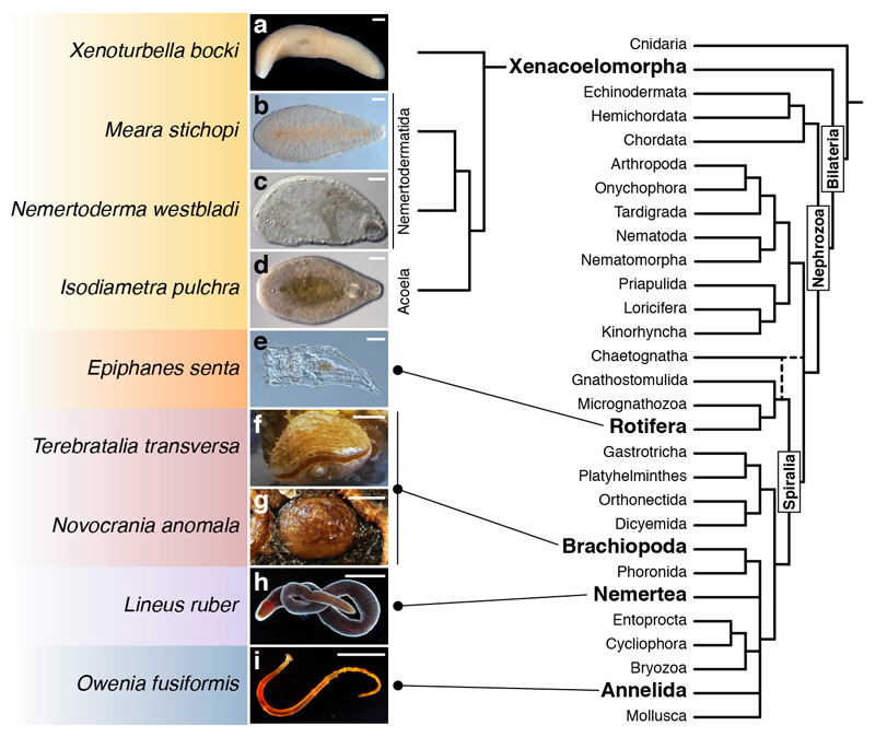
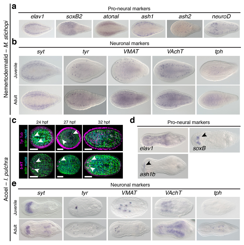
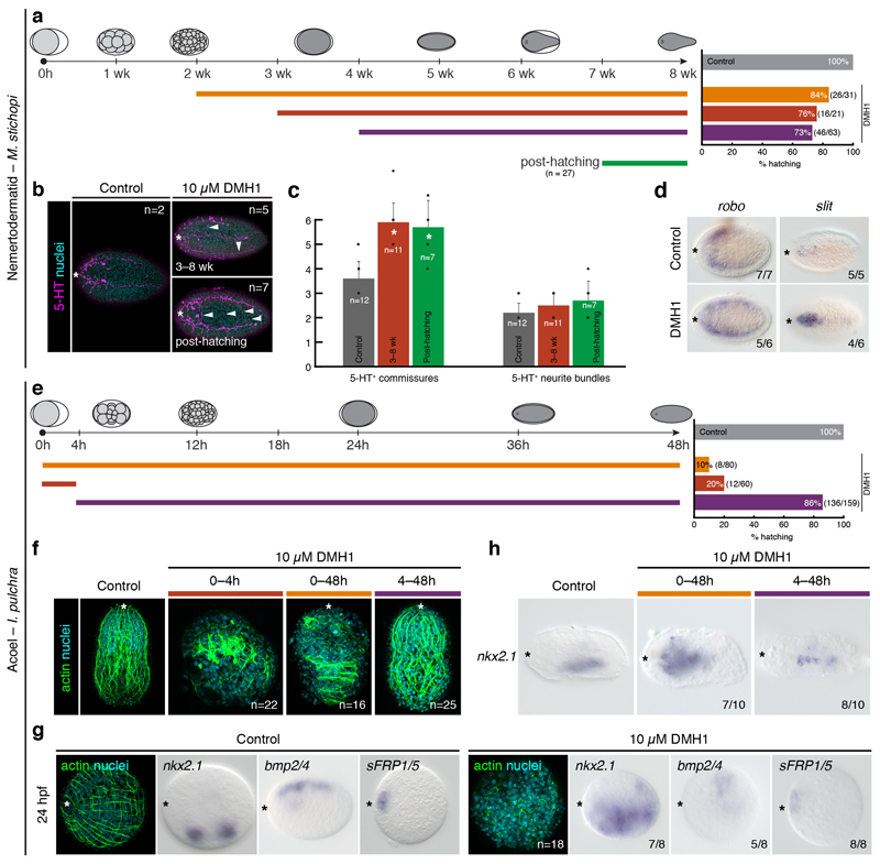
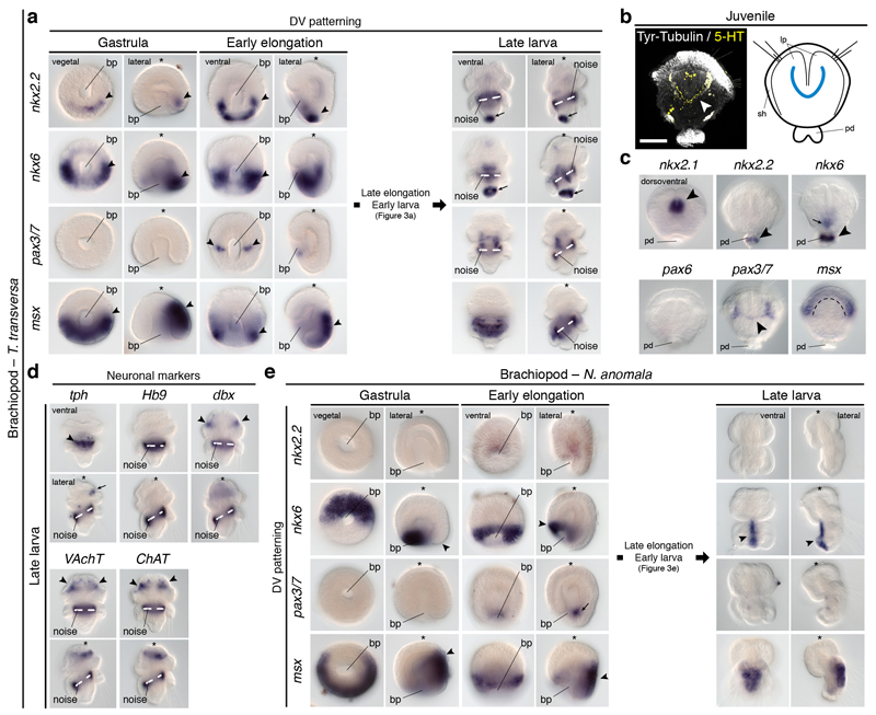
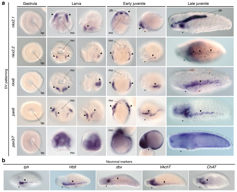
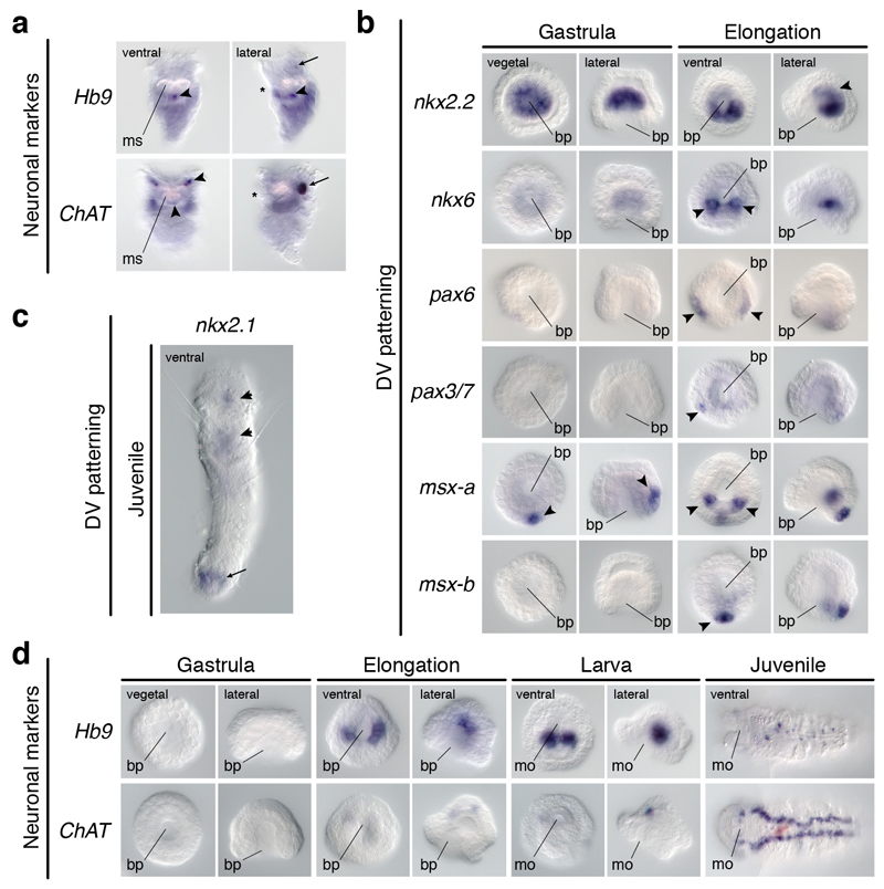
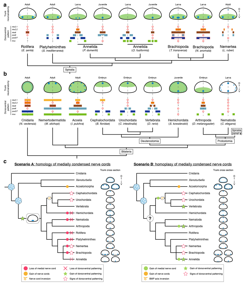
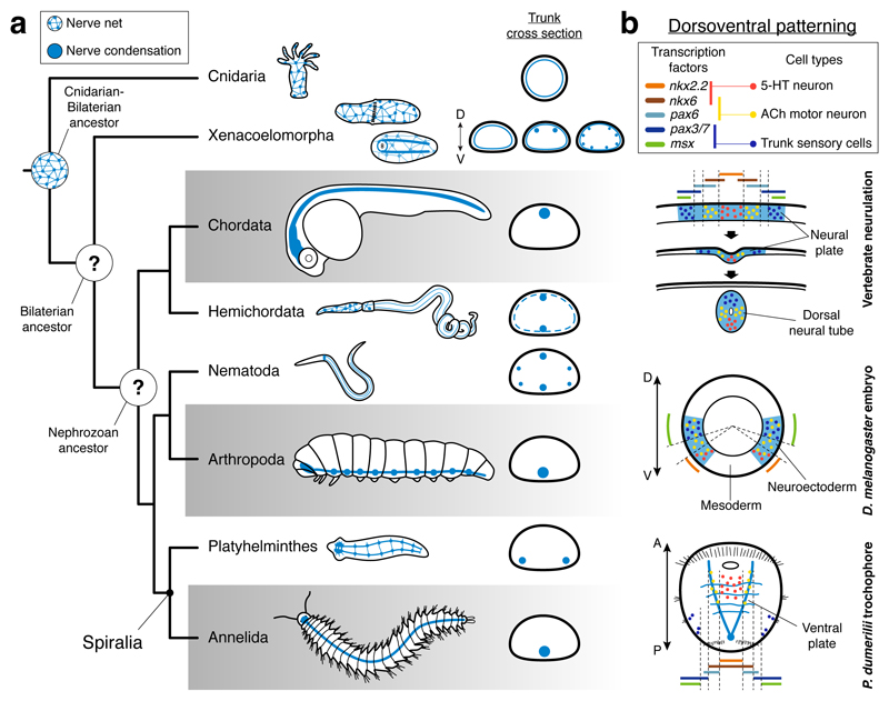
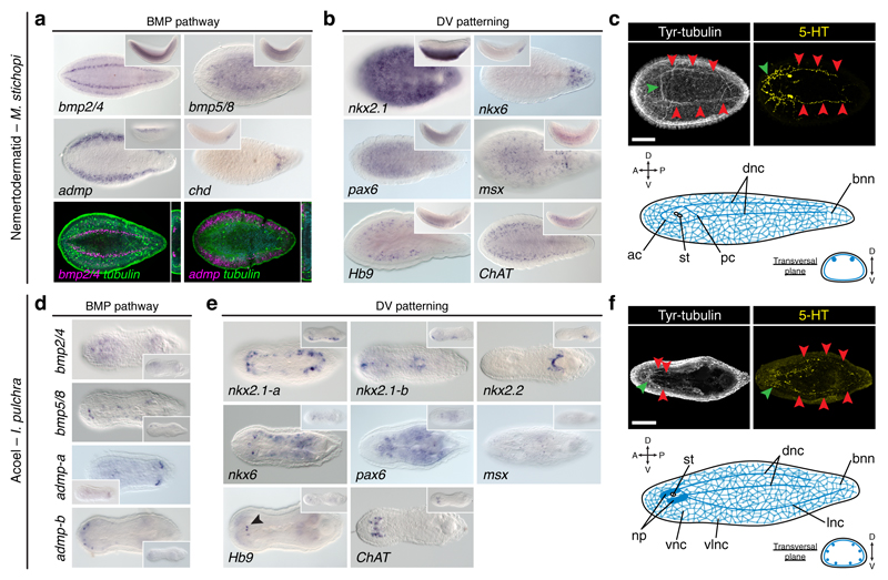
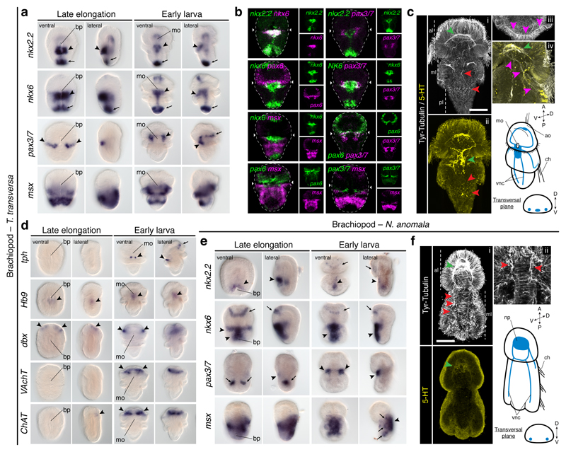
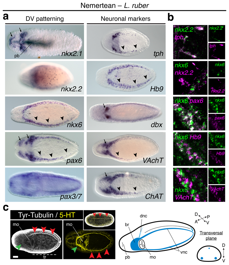
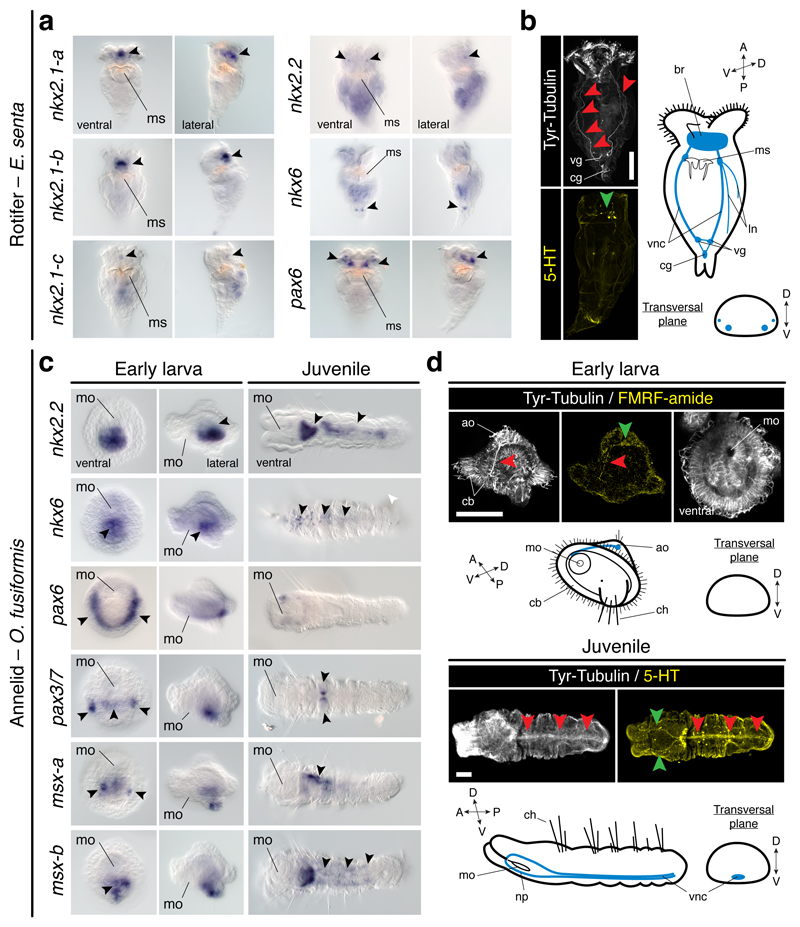
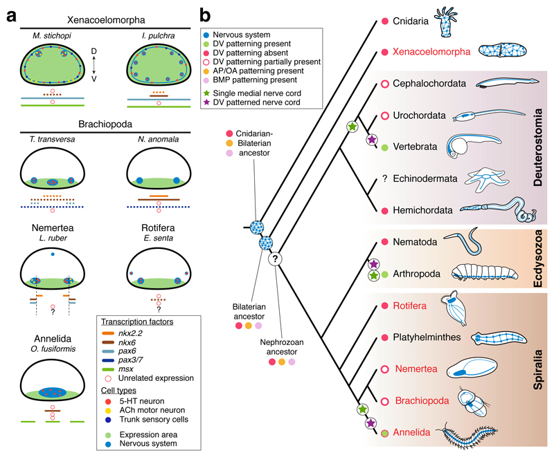
It has been hypothesized that a condensed nervous system with a medial ventral nerve cord is an ancestral character of Bilateria. The presence of similar dorsoventral molecular patterns along the nerve cords of vertebrates, flies, and an annelid has been interpreted as support for this scenario. Whether these similarities are generally found across the diversity of bilaterian neuroanatomies is unclear, and thus the evolutionary history of the nervous system is still contentious. Here we study representatives of Xenacoelomorpha, Rotifera, Nemertea, Brachiopoda, and Annelida to assess the conservation of the dorsoventral nerve cord patterning. None of the studied species show a conserved dorsoventral molecular regionalization of their nerve cords, not even the annelid Owenia fusiformis, whose trunk neuroanatomy parallels that of vertebrates and flies. Our findings restrict the use of molecular patterns to explain nervous system evolution, and suggest that the similarities in dorsoventral patterning and trunk neuroanatomies evolved independently in Bilateria.


Conflict of interest statement

The authors declare no competing financial interests.

Figures

Extended Data Figure 1
Extended Data Figure 1. Studied species.
(ai) Images of the adult forms of the studied species within a consensus bilaterian phylogeny. Color boxes highlight major taxonomical clades. Scale bars, 100 µm in ae, 0.5 cm in h and g, 1 cm in f, h, and i.
Extended Data Figure 2
Extended Data Figure 2. Gene expression in Xenoturbella bocki and Nemertoderma westbladi.
(a) Two six3/6 paralogs are expressed in the anterior head margin in X. bocki (arrowheads). (b) The neural marker synaptotagmin (syt) is detected in the circumferential (cf; inset 1) and side (sf; inset 2) sensory furrows in X. bocki. (c) In N. westbladi, the anterior marker sFRP1/5 (arrowhead) and the posterior genes gbx and wnt1 are asymmetrically expressed along the anteroposterior axis of N. westbladi. (d) The BMP ligands bmp2/4-a and bmp2/4-c are expressed dorsally, while the BMP antagonist admp is expressed dorsolaterally. (e) The CNS of N. westbladi comprises an anterior ring-like commissure (green arrowheads) and a main pair of ventral condensations (red arrowhead). (f) The neuronal marker syt is highly expressed in the anterior part (inset 1), and in the nerve cords (inset 2). In the different panels, dotted rectangles indicate magnified areas. In all panels, the anterior pole is to the left. The schematic drawing in c is not to scale. Scale bar, 100 µm in c.
Extended Data Figure 3
Extended Data Figure 3. Anteroposterior patterning in Xenacoelomorpha.
(a, b) Expression of anteroposterior markers in adult specimens of M. stichopi and I. pulchra. In both species, sFRP1/5, vax, six3/6, and BarH are expressed in anterior territories (black arrowheads). In M. stichopi, Rx is also expressed anteriorly, but broadly along the animal body in I. pulchra. In this acoel, emx is detected in the anterior part of the animal (background staining close to the gonads). In the nemertodermatid, the anterior neural markers otx, otp, pax2/5/8, and fezf are expressed along the entire AP axis, in association with the dorsal nerve cords (black dotted lines in otp). In I. pulchra, otx, pax2/5/8-a, and pax2/5/8-b are broadly expressed. In M. stichopi, an irx ortholog is detected in the posterior tip, while it is detected in the anterior tip and around the mouth and copulatory apparatus in the acoel (arrowheads). The gbx ortholog of M. stichopi is expressed posteriorly, and the trunk-related Hox genes are expressed in two lateral rows (anterior Hox) and anteriorly to the mouth and in the posterior tip (posterior Hox). In the nemertodermatid and the acoel, Wnt ligand genes are expressed posteriorly (arrowheads). All images are dorsoventral views with anterior to the left. (c) Schematic summary of anteroposterior expression in the nemertodermatid M. stichopi and the acoel I. pulchra. Drawings are not to scale and the extent of the expression domains are only approximate. The expression of posterior Hox in I. pulchra is based on.
Extended Data Figure 4
Extended Data Figure 4. Expression of BMP components in Nemertodermatida and Acoela.
(a) In the nemertodermatid Meara stichopi, the BMP pathway antagonists twisted gastrulation (tsg), and crossveinless 2 (cv2) are expressed dorsally, while the antagonist BAMBI is broadly detected in the ventral side. The gene tolloid (tld) is expressed both dorsally and ventrally. The BMP receptors bmpR-I is expressed dorsolaterally and the bmpR-II is detected more broadly. The genes smad1 and smad4 are expressed broadly and smad6 is expressed along the dorsal nerve cords. (b) bmp2/4 is not expressed in neuronal cells (elav1+ cells), but in cells located medially to the nerve cords (tubulin positive). The cells expressing bmp2/4 also express tsg, and cv2 is expressed dorsally along the nerve cords. (c) In the acoel Isodiametra pulchra, the BMP antagonist tld is expressed ventrally, the bmpR-I is detected in the inner body, and bmpR-II is expressed anteriorly and posteriorly around the copulatory organ. The genes smad1 and smad4 are expressed generally, while smad6 is expressed in two bilaterally symmetrical anterior clusters. All main panels are dorsoventral views, and the insets are lateral views.
Extended Data Figure 5
Extended Data Figure 5. Expression of neuronal markers in Nemertodermatida and Acoela.
(a) In the nemertodermatid Meara stichopi, the genes associated with neuronal fate commitment elav1, soxB2, ash1, ash2, atonal, and neuroD are detected along the dorsal nerve cords. (b) Similarly, the neuronal markers synaptotagmin (syt), tyrosine hydroxylase (tyr), vesicular monoamine transporter (VMAT), choline acetyltransferase (ChAT), vesicular acetylcholine transporter (VAchT), and tryptophan hydroxylase (tph) are mostly expressed dorsally, along the dorsal nerve cords. (c) Morphology of I. pulchra embryos stained against tyrosinated tubulin (Tyr tub) and serotonin (5-HT), and counterstained with phallacidin (actin bundles) and DAPI (nuclei). The first tubulin-positive cells that resemble neurons appear anteriorly (arrowheads) at 24 hours post-fertilization (hpf). By 32 hpf, the anterior and posterior lobes of the brain, as well as some neurite bundles are visible. Similarly, the first serotonergic cells are detected at 24 hpf in the anterior end (arrowheads). (d) In I. pulchra, the pro-neural marker elav1 is broadly expressed, soxB is detected in the head region (arrowhead), and ash1b in the anterior tip (arrowhead). (e) In I. pulchra, the neuronal marker syt is highly expressed in the anterior neuropile. The marker tyr is detected in the statocyst and isolated cells. VMAT is detected in isolated dorsal cell clusters in the juvenile that concentrate along the adult brain. ChAT and VAchT are expressed in the brain in juveniles and adults (gonadal staining in the adult is background). The gene tph is expressed in isolated ventral cells of the adult. All panels are dorsoventral views with anterior to the left. Scale bars, 50 µm in c.
Extended Data Figure 6
Extended Data Figure 6. DMH1 treatments in Meara stichopi and Isodiametra pulchra.
(a) Schematic overview of dorsomorphin homologue 1 (DMH1) treatments in M. stichopi and % of hatching embryos for each experimental condition. (b) M. stichopi embryos incubated with DMH1 from 3 to 8 weeks and after hatching show more serotonergic commissures that control animals. (c) The differences in the number of commissures are significant in both pre-hatching (asterisk; two-tail t-test; p-value < 0.0001) and post-hatching (asterisk; two-tail t-test; p-value < 0.0014) treated embryos. In contrast, the number of serotonin positive neurite bundles is not significantly increased in any of the treatments. (d) Despite the abnormal development of serotonergic axonal tracts, slit and robo genes are expressed similarly. The differences in signal intensity are due to technical variability. (e) Schematic overview of DMH1 treatments in I. pulchra and % of hatching embryos for each experimental condition. (f) Morphological analyses of DMH1 treated embryos. Treatment in early stages affects normal development, while treatments from 4 hours onwards does not significantly compromises embryogenesis. (g) Embryos treated between 0–4 hours post fertilization and fixed at 24 hours of development show expanded expression of the ventral marker nkx2.1, reduced expression of the dorsal gene bmp2/4, and unaffected expression of the anterior marker sFRP1/5. The embryo shows a disorganized morphology, as revealed by actin staining. (h) The expression of the ventral marker nkx2.1 is expanded in early treated embryos (0–48 h), but unaffected in embryos treated after 4 hours of development. In (b, d, fh), the asterisk marks the anterior pole. In (b, d, f), panels are dorsoventral views, and in (g, h) the panels are lateral views.
Extended Data Figure 7
Extended Data Figure 7. Gene expression in Brachiopoda.
(a) Gene expression during early gastrulation and elongation, and in late larvae of T. transversa. The gene nkx2.2 is expressed ventroposteriorly (black arrowhead) and in the pedicle lobe of the larva (arrow). nkx6 is detected in two bilateral symmetrical ectodermal posterior clusters (arrowheads) and in the archenteron wall. In the larva, nkx6 is expressed in the pedicle lobe (arrow) and midgut. pax3/7 is first detected in two ventrolateral domains at the prospective apical-trunk boundary (arrowheads), and in the ventral anterior region of the larva. The gene msx is first expressed dorsally, in the future mantle ectoderm (arrowheads), and in the mantle of the larva. (b) In 2-days old post-metamorphic juveniles, the CNS comprises a main serotonergic anterior commissure (white arrowhead; dorsoventral view) that innervates the developing lophophore. The schematic drawing is not to scale, and the blue line represent the commissure. (c) The gene nkx2.1 is expressed in the anterior region (arrowhead), between the lophophores in 2-day old juveniles. The genes nkx2.2 and nkx6 are expressed in the pedicle (arrowheads), and nkx6 is also detected in the gut (arrow). The gene pax6 shows no expression, pax3/7 is detected in the neural commissure (arrowhead), and msx is expressed in the cells at the edge of the mantle (dotted line). (d) Neuronal markers in late larvae of T. transversa. The serotonergic marker tph is expressed in the anteroventral condensation of the mantle lobe (arrowhead) and in dorsal ectodermal cells of the apical lobe (arrow). No expression is detected for Hb9, and the genes dbx, VAchT and ChAT are all detected in the anterior apical neuroectoderm (arrowheads). (e) Gene expression during early gastrulation and elongation, and in late larvae of N. anomala. The gene nkx2.2 is expressed in the anterior blastoporal lip at the onset of axial elongation, and it is not detected in the late larva. nkx6 is asymmetrically expressed around the blastopore, in the putative anteroventral ectoderm (arrowhead). As the blastopore closes, the expression extends posteriorly and concentrates along the midline of the larva (arrowhead). The gene pax3/7 is detected in the posterior mesoderm at the onset of axial elongation (arrow). The gene msx is expressed in the prospective mantle lobe ectoderm (arrowheads) and in the dorsal shell-forming epithelium of the late larva. The asterisks indicate the animal/anterior pole and white dotted lines in (a, d) mark the region of background noise caused by probe trapping in the shell forming ectoderm. Panel orientations are indicated in the first row/column and apply to the rest of the panels in the same column/row. Scale bar, 100 µm in b.
Extended Data Figure 8
Extended Data Figure 8. Gene expression in the nemertean Lineus ruber.
(a) None of the nerve cord patterning genes is expressed during gastrulation in L. ruber. In the intracapsular larva, nkx2.1 is expressed in the cephalic imaginal discs (arrowheads), nkx2.2 and nkx6 in an anterior and a posterior domain of the trunk imaginal discs (arrowheads) respectively, and pax6 is detected both in the cephalic and the anterior trunk imaginal discs (arrowheads). pax3/7 is broadly expressed. With metamorphosis, nkx2.1 is detected in the head and proboscis, nkx2.2 is detected in the nerve cords and isolated trunk cells (arrowheads), nkx6 is expressed in the nerve cords (arrowheads), pax6 is observed in the head and nerve cords (arrowheads), and pax3/7 remains broadly expressed. All gastrulae are vegetal views. For larvae and early juveniles, the left column is a dorsoventral view and the right column is a lateral view (anterior to the left). All late juvenile pictures are lateral views, with anterior to the left. (b) Lateral views (anterior to the left) of neuronal markers in juveniles. They are all expressed in the ventral nerve cords (arrowheads), and not in the dorsal neurite bundle. In all panels, the asterisk indicates the position of the mouth opening. bp, blastopore; mo, mouth; pb, proboscis.
Extended Data Figure 9
Extended Data Figure 9. Molecular patterning and motoneuron markers in Rotifera and Annelida.
(a) Expression of the motoneuron markers Hb9 and ChAT in juveniles of the rotifer Epiphanes senta. The gene Hb9 is detected in neurons of the mastax (arrowheads) and weakly in isolated cells of the brain (arrow). The gene ChAT is detected in the brain (arrow), cells of the corona and mastax (arrowheads). (b) Expression of dorsoventral patterning genes in gastrulae and elongating embryos of Owenia fusiformis. nkx2.2 and nkx6 are expressed in the internalized endomesoderm (arrowheads). The gene pax6 is expressed in two lateral rows during elongation (arrowhead) and pax3/7 in two lateral cells (arrowhead). Of the two paralogs, msx-a is first detected in a posterior ectodermal domain (arrowhead) and in two additional bilaterally symmetrical posterior cells (arrowheads) during elongation. The gene msx-b is only detected during elongation in a posterior domain (arrowhead). (c) Ventral view of the expression of nkx2.1 in the juvenile of the annelid O. fusiformis. This gene is detected in the foregut (arrowheads) and hindgut (arrow). (d) Expression of the motoneuron markers Hb9 and ChAT in O. fusiformis. Hb9 is first detected in lateral domains of the archenteron/gut during embryogenesis and larva, and in isolated cells of the ventral trunk of the juvenile. The gene ChAT is detected in three cells of the apical region of the embryo and larva, and in the neuropile and two lateral ventral cords of the juvenile. bp, blastopore; mo, mouth; ms, mastax. The asterisk in (a) marks the position of the mouth.
Extended Data Figure 10
Extended Data Figure 10. Dorsoventral patterning and the evolution of bilaterian trunk neuroanatomy.
(a, b) Schematic drawings of trunk neuroanatomy (nerve cords in blue) and expression of patterning genes in spiralian (a) and bilaterian (b) lineages. The overall location of patterning genes expression domains with respect to the DV axis and nerve cords is indicated by light green. In (a), the red dashed squared expression of pax6 and pax3/7 in brachiopods indicates that these expression domains are only in the anterior region of the mantle lobe, not all along the trunk. Similarly, the red dashed squared expression of nkx6 in rotifers highlights that this gene is only expressed posteriorly in the trunk. In (b), the red dashed squared expression of nkx2.1, nkx2.2 and nkx6 in Cnidaria indicates that these genes are expressed in the pharynx ectoderm. The red dashed squared expression of nkx6 in M. stichopi shows that this gene is only expressed posteriorly. In the acoel I. pulchra, the red dashed squared expression of nkx2.2 specifies that this gene is only expressed between mouth and copulatory organ. Red circles imply that a gene is not expressed in the trunk or is missing. Question marks indicate that there is no available data about the expression of that particular gene. See Extended Data Table 1 and main text for references. Schematic drawings are not to scale and only represent approximate relative expression domains. (c) Alternative scenarios for the evolution of the dorsoventral patterning and bilaterian nerve cords. In scenario A, the medially condensed nerve cords of vertebrates, arthropods, and annelids are homologous. Therefore, the dorsoventral patterning was lost multiple times both in lineages with medially condensed nerve cords (e.g. the annelid Owenia fusiformis, cephalochordates, and tunicates) and in lineages with multiple nerve cords and diffuse nerve nets. In scenario B, which is supported by this study and is more parsimonious, the similarities in dorsoventral patterning and trunk neuroanatomies of vertebrates, arthropods, and some annelids evolved convergently. The diversity of nerve cord arrangements in nephrozoan lineages hampers reconstructing the ancestral neuroanatomy for this group (question mark). Animal phylogeny based on
Figure 1
Figure 1. CNS evolution and dorsoventral patterning.
(a) A nerve net is ancestral for Cnidaria and Bilateria. The neuroanatomical diversity hampers the reconstruction of the CNS evolution in Bilateria. (b) A central argument for an ancestral medially condensed VNC for Bilateria is the similar deployment of DV-TFs in vertebrates, Drosophila, and the P. dumerilii larva. The staggered expression of these genes concurs with specific neuronal populations.
Figure 2
Figure 2. Dorsoventral patterning in Xenacoelomorpha.
(a) bmp ligands and admp are expressed dorsally; chd is expressed ventroposteriorly. (b) nkx2.1, nkx6, and msx are expressed ventrally; pax6 is expressed broadly; Hb9 and ChAT are in the nerve cords. (c) M. stichopi CNS (green arrowheads indicate the anterior commissures; red arrowheads indicate the nerve cords). (d) bmp ligands are expressed dorsally; admp-a is expressed posteroventrally; admp-b is expressed anterolaterally. (e) nkx2.1 paralogs and nkx2.2 are expressed ventrally; nkx6 is expressed laterally; pax6 throughout the body; msx in isolated cells; Hb9 and ChAT are in the brain. (f) I. pulchra CNS (green arrowheads indicate the brain; red arrowheads indicate the nerve cords). Insets are lateral views. ac, anterior commissure; bnn, basiepidermal nerve net; dnc, dorsal nerve cord; lnc, lateral nerve cord; np, neuropile; pc, posterior commissure; st, statocyst; vlnc, ventrolateral nerve cord; vnc, ventral nerve cord. Scale bars, 100 µm.
Figure 3
Figure 3. Dorsoventral patterning in Brachiopoda.
(a) nkx2.2 and nkx6 are in the trunk midline (arrowheads), posterior tip (arrows), gut, and apical cells (nkx6); pax3/7 is expressed laterally (arrowheads) and in the apical lobe (arrow); msx is in the mantle and ventral pedicle. (b) There is an nkx2.2+/nkx6+ medioventral region, and a more lateral nkx6+/pax6+/pax3/7+ anterior trunk domain. (c) T. transversa larval CNS (green arrowheads indicate the neuropile in i, and the trunk serotonergic condensation in ii; red arrowheads mark the VNCs; pink arrowheads indicate the innervation of the chaetae). (d) Only tph is expressed in the trunk (arrows and arrowheads indicate expression areas). (e) nkx2.2 and nkx6 are in the trunk ventral midline (arrowheads), apical lobe (arrows), and gut; pax3/7 is in the mesoderm (arrows), and in two ventrolateral trunk domains (arrowheads); msx is in the trunk, shell epithelium (arrowhead), and mesoderm (arrows). (f) N. anomala larval CNS (green arrowhead indicates the neuropile; red arrowheads in i mark the VNCs; red arrowheads in ii indicate the innervation of the chaetae). ao, apical organ; bp, blastopore; ch, chaetae; mo, mouth; np, neuropile; vnc, ventral nerve cord. Scale bars, 50 µm.
Figure 4
Figure 4. Dorsoventral patterning in Nemertea.
(a) nkx2.1 and nkx2.2 are in the head (arrows), proboscis (nkx2.1), and trunk cells (nkx2.2); nkx6 and pax6 are in the head (arrows) and VNCs (arrowheads); pax3/7 is broadly expressed. Neuronal markers are in the brain (arrows) and VNCs (arrowheads). (b) In the VNCs, nkx2.2+ cells express tph, but not nkx6; nkx6+ cells express pax3/7 and Hb9, but not VAchT. (c) L. ruber CNS (green arrowheads indicate the brain; red arrowheads mark the VNCs and the dorsal neurite in the upper inset). br, brain; dnc, dorsal nerve cord; mo, mouth; tr, trunk; pb, proboscis; vnc, ventral nerve cord. Scale bar, 100 µm.
Figure 5
Figure 5. Dorsoventral patterning in Rotifera and Annelida.
(a) nkx2.1 paralogs, nkx2.2, and pax6 show brain domains (arrowheads); nkx6 is detected posteriorly (arrowheads). (b) E. senta CNS (green arrowhead indicates the brain; red arrowheads indicate the VNCs, and additional neurites). (c) nkx2.2 shows gut expression (arrowhead); nkx6 is in the ventral midline (arrowheads); pax6 is in two lateral larval bands (arrowheads), and juvenile head; pax3/7 is in two ventrolateral larval clusters and midline (arrowheads), but in two trunk clusters in juveniles (arrowheads); msx paralogs are in ventral larval domains and the juvenile VNC (arrowheads). (d) O. fusiformis CNS (green arrowheads indicate the apical larval FMRF-amide cell and juvenile brain; red arrowheads indicate the larval anterior axon and juvenile medial VNC). ao, apical organ; br, brain; cb, ciliary band; cg, caudal ganglion; ch, chaetae; ln, lateral neurites; mo, mouth; ms, mastax; np, neuropile; vg, vesicle ganglia; vnc, ventral nerve cord. Scale bars, 50 µm.
Figure 6
Figure 6. Dorsoventral patterning and CNS evolution.
(a) Gene expression summary. Dotted lines indicate that the expression does not extend along the entire trunk. (b) Proposed scenario for the evolution of neuroectodermal patterning systems in Bilateria. A nerve net, and the anteroposterior (AP) and BMP axial patterning predate the Cnidaria-Bilateria split, and were present in the Bilateria ancestor. The ancestral nephrozoan neuroanatomy remains unclear (question mark). The dorsoventral (DV) patterning system is not tied to the CNS arrangement in Bilateria (as in Chordata and Annelida). In red, lineages analysed in this study. The green dot with red border indicates that there are annelids with and without the DV patterning.

Comment in

References

    1. Schmidt-Rhaesa A, Harzsch S, Purschke G. Structure & Evolution of Invertebrate Nervous Systems. Oxford University Press; 2016.
    1. Arendt D, Tosches MA, Marlow H. From nerve net to nerve ring, nerve cord and brain--evolution of the nervous system. Nat Rev Neurosci. 2016;17:61–72. - PubMed
    1. Hejnol A, Pang K. Xenacoelomorpha's significance for understanding bilaterian evolution. Curr Opin Genet Dev. 2016;39:48–54. - PubMed
    1. Arendt D, Denes AS, Jékely G, Tessmar-Raible K. The evolution of nervous system centralization. Philos Trans R Soc Lond B Biol Sci. 2008;363:1523–1528. - PMC - PubMed
    1. Hejnol A, Lowe CJ. Embracing the comparative approach: how robust phylogenies and broader developmental sampling impacts the understanding of nervous system evolution. Phil Trans R Soc B. 2015;370 20150045. - PMC - PubMed

Publication types