Comparison between conventional protective mechanical ventilation and high-frequency oscillatory ventilation associated with the prone position
- PMID: 29236845
- PMCID: PMC5764554
- DOI: 10.5935/0103-507X.20170067
Comparison between conventional protective mechanical ventilation and high-frequency oscillatory ventilation associated with the prone position
Abstract
Objective: To compare the effects of high-frequency oscillatory ventilation and conventional protective mechanical ventilation associated with the prone position on oxygenation, histology and pulmonary oxidative damage in an experimental model of acute lung injury.
Methods: Forty-five rabbits with tracheostomy and vascular access were underwent mechanical ventilation. Acute lung injury was induced by tracheal infusion of warm saline. Three experimental groups were formed: healthy animals + conventional protective mechanical ventilation, supine position (Control Group; n = 15); animals with acute lung injury + conventional protective mechanical ventilation, prone position (CMVG; n = 15); and animals with acute lung injury + high-frequency oscillatory ventilation, prone position (HFOG; n = 15). Ten minutes after the beginning of the specific ventilation of each group, arterial gasometry was collected, with this timepoint being called time zero, after which the animal was placed in prone position and remained in this position for 4 hours. Oxidative stress was evaluated by the total antioxidant performance assay. Pulmonary tissue injury was determined by histopathological score. The level of significance was 5%.
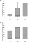
Results: Both groups with acute lung injury showed worsening of oxygenation after induction of injury compared with the Control Group. After 4 hours, there was a significant improvement in oxygenation in the HFOG group compared with CMVG. Analysis of total antioxidant performance in plasma showed greater protection in HFOG. HFOG had a lower histopathological lesion score in lung tissue than CMVG.
Conclusion: High-frequency oscillatory ventilation, associated with prone position, improves oxygenation and attenuates oxidative damage and histopathological lung injury compared with conventional protective mechanical ventilation.
Objetivo: Comparar os efeitos da ventilação oscilatória de alta frequência e da ventilação mecânica convencional protetora associadas à posição prona quanto à oxigenação, à histologia e ao dano oxidativo pulmonar em modelo experimental de lesão pulmonar aguda.
Métodos: Foram instrumentados com traqueostomia, acessos vasculares e ventilados mecanicamente 45 coelhos. A lesão pulmonar aguda foi induzida por infusão traqueal de salina aquecida. Foram formados três grupos experimentais: animais sadios + ventilação mecânica convencional protetora, em posição supina (Grupo Controle; n = 15); animais com lesão pulmonar aguda + ventilação mecânica convencional protetora, posição prona (GVMC; n = 15); animais com lesão pulmonar aguda + ventilação oscilatória de alta frequência, posição prona (GVAF; n = 15). Após 10 minutos do início da ventilação específica de cada grupo, foi coletada gasometria arterial, sendo este momento denominado tempo zero, após o qual o animal foi colocado em posição prona, permanecendo assim por 4 horas. O estresse oxidativo foi avaliado pelo método de capacidade antioxidante total. A lesão tecidual pulmonar foi determinada por escore histopatológico. O nível de significância adotado foi de 5%.
Resultados: Ambos os grupos com lesão pulmonar aguda apresentaram piora da oxigenação após a indução da lesão comparados ao Grupo Controle. Após 4 horas, houve melhora significante da oxigenação no grupo GVAF comparado ao GVMC. A análise da capacidade antioxidante total no plasma mostrou maior proteção no GVAF. O GVAF apresentou menor escore de lesão histopatológica no tecido pulmonar que o GVMC.
Conclusão: A ventilação oscilatória de alta frequência, associada à posição prona, melhora a oxigenação, e atenua o dano oxidativo e a lesão pulmonar histopatológica, comparada com ventilação mecânica convencional protetora.
Conflict of interest statement
Figures


References
-
- ARDS Definition Task Force. Ranieri VM, Rubenfeld GD, Thompson BT, Ferguson ND, Caldwell E, Fan E, et al. Acute respiratory distress syndrome. The Berlin Definition. JAMA. 2012;307(23):2526–2533. - PubMed
-
- Arnold JH, Hanson JH, Toro-Figuero LO, Gutiérrez J, Berens RJ, Anglin DL. Prospective, randomized comparison of high-frequency oscillatory ventilation and conventional ventilation in pediatric respiratory failure. Crit Care Med. 1994;22(10):1530–1539. - PubMed
-
- Froese AB, McCulloch PR, Sugiura M, Vaclavik S, Possmayer F, Moller F. Optimizing alveolar expansion prolongs the effectiveness of exogenous surfactant therapy in the adult rabbit. Am Rev Respir Dis. 1993;148(3):569–577. - PubMed
-
- Fioretto JR, Rebello CM. High frequency oscillatory ventilation in pediatrics and neonatology. Rev Bras Ter Intensiva. 2009;21(1):96–103. - PubMed
Publication types
MeSH terms
Substances
LinkOut - more resources
Full Text Sources
Other Literature Sources

