. 2018 Mar 1;114(3):417-430.
doi: 10.1093/cvr/cvx242.

Impaired cardiac contractile function in arginine:glycine amidinotransferase knockout mice devoid of creatine is rescued by homoarginine but not creatine

Affiliations

Impaired cardiac contractile function in arginine:glycine amidinotransferase knockout mice devoid of creatine is rescued by homoarginine but not creatine

Kiterie M E Faller et al. Cardiovasc Res. .

Abstract

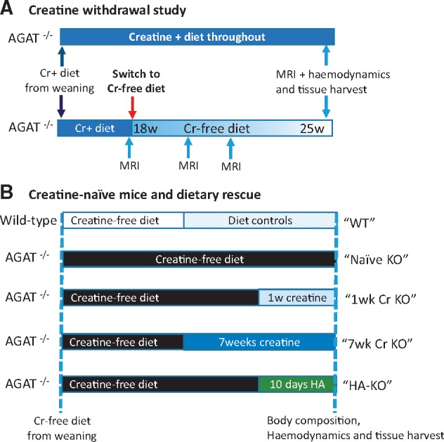
Aims: Creatine buffers cellular adenosine triphosphate (ATP) via the creatine kinase reaction. Creatine levels are reduced in heart failure, but their contribution to pathophysiology is unclear. Arginine:glycine amidinotransferase (AGAT) in the kidney catalyses both the first step in creatine biosynthesis as well as homoarginine (HA) synthesis. AGAT-/- mice fed a creatine-free diet have a whole body creatine-deficiency. We hypothesized that AGAT-/- mice would develop cardiac dysfunction and rescue by dietary creatine would imply causality.

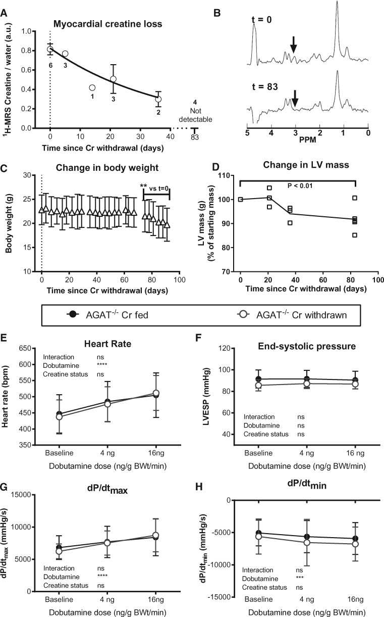
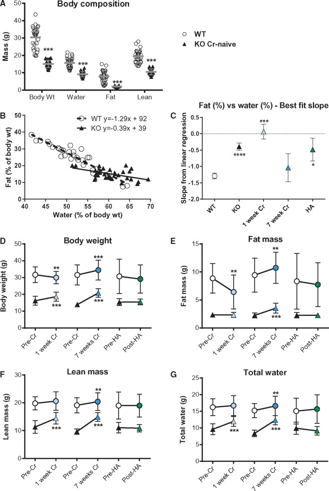
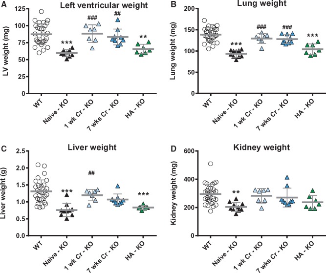
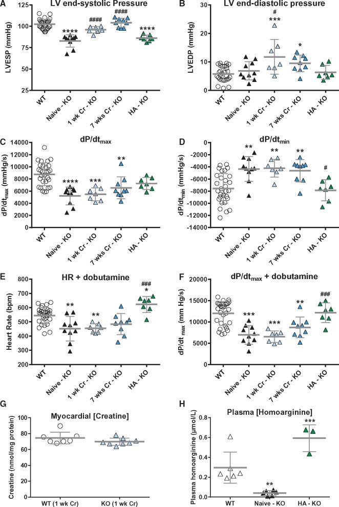
Methods and results: Withdrawal of dietary creatine in AGAT-/- mice provided an estimate of myocardial creatine efflux of ∼2.7%/day; however, in vivo cardiac function was maintained despite low levels of myocardial creatine. Using AGAT-/- mice naïve to dietary creatine we confirmed absence of phosphocreatine in the heart, but crucially, ATP levels were unchanged. Potential compensatory adaptations were absent, AMPK was not activated and respiration in isolated mitochondria was normal. AGAT-/- mice had rescuable changes in body water and organ weights suggesting a role for creatine as a compatible osmolyte. Creatine-naïve AGAT-/- mice had haemodynamic impairment with low LV systolic pressure and reduced inotropy, lusitropy, and contractile reserve. Creatine supplementation only corrected systolic pressure despite normalization of myocardial creatine. AGAT-/- mice had low plasma HA and supplementation completely rescued all other haemodynamic parameters. Contractile dysfunction in AGAT-/- was confirmed in Langendorff perfused hearts and in creatine-replete isolated cardiomyocytes, indicating that HA is necessary for normal cardiac function.

Conclusions: Our findings argue against low myocardial creatine per se as a major contributor to cardiac dysfunction. Conversely, we show that HA deficiency can impair cardiac function, which may explain why low HA is an independent risk factor for multiple cardiovascular diseases.


Figures

Figure 1
Figure 1
Schematic showing experimental study design. (A) Creatine withdrawal study: AGAT-/- mice were bred and weaned onto a diet containing 0.5% creatine (w/w), which was then switched to a standard creatine-free diet at 18 weeks of age in the experimental group. These animals received multiple MRI and 1H-MRS examinations before LV haemodynamics and tissue harvest at ∼25 weeks of age. (B) Creatine-naïve mice and dietary rescue: WT and AGAT-/- mice were bred and weaned onto a standard creatine-free diet for the first 4–5 months of life. Mice were then either maintained on this creatine-free diet, switched to a creatine supplemented diet for either 1 or 7 weeks, or given 14 mg/L of homoarginine for 10 days while maintaining a creatine-free diet. Wild-type controls were included for all dietary manipulations.
Figure 2
Figure 2
Withdrawal of dietary creatine in AGAT-/- mice reduces body weight and LV mass without affecting haemodynamic function. (A) Myocardial creatine depletion was estimated at 2.7 ± 0.4% of the free creatine pool per day using in vivo1H-MRS. Data were fitted using a kinetic model of non-enzymatic degradation, according to the following equation: [Cr]t = [Cr]t=0. e-kt. (B) Representative 1H-MRS spectra of the same mouse before and after creatine withdrawal. The creatine peak (arrow) seen at day 0 is not visible by day 83. (C) Body weight decreased rapidly after 70 days of creatine-free diet (n = 6). (D) LV mass calculated by in vivo cine-MRI falls during dietary creatine withdrawal. LV haemodynamic parameters were measured at day 90 in AGAT-/- mice with and without dietary creatine withdrawal (n = 6/group) under resting baseline conditions and with IV infusion of dobutamine. There were no significant differences between groups for (E) heart rate, (F) LV end-systolic pressure, (G) the rate of pressure rise maximum (dP/dtmax) as a measure of contractility, or (H) the rate of pressure rise minimum (dP/dtmin) as a measure of relaxation. Comparison was made by two-way repeated measures ANOVA and data are represented as mean ± SD.
Figure 3
Figure 3
Absence of phosphocreatine (PCr) in creatine-naïve AGAT knockout mice does not alter key metabolic parameters in the heart. Representative 31P-magnetic resonance spectra in Langendorff-perfused hearts. PCr was the most prominent peak in hearts from wild-type mice (A), but was completely absent in hearts from creatine-naïve AGAT-/- mice (B), where inorganic phosphate (Pi) was elevated, and there was no change in ATP (appears as three peaks representing the γ, α, and β phosphoryl groups). Key energy homeostasis enzymes and mitochondrial function were not significantly different between wild-type (WT) and creatine-naïve AGAT-/- (KO) hearts. (C) Total creatine kinase (CK) activity and percentage isoenzyme distribution (D), where Mito is mitochondrial CK and the various dimers of Muscle and Brain isoforms; (E) adenylate kinase (AK) activity (all n = 10 WT, n = 13 KO). (F) AMPK activation expressed as the ratio of phospho- to total AMPK protein expression was not altered in LV, but was significantly elevated in hind-limb skeletal muscle, n = 5 per group. (G) Citrate synthase activity (n = 10 WT, n = 13 KO) and (H) mitochondrial respiration with glutamate (5 mM), malate (2.5 mM) and Na+-pyruvate (5 mM) as substrates (n = 7 WT, n = 8 KO). Values are mean ± SD.
Figure 4
Figure 4
Creatine-naïve AGAT-/- mice have low body weight and altered composition rescuable by dietary creatine. (A) Creatine-naïve KO mice had low body weight associated with reduced water, fat and lean mass (***P < 0.001; n = 14 male and 14 female per group). (B) These changes were not proportional because the linear relationship between % body fat and % total water was significantly altered in KO mice (P < 0.0001 for slope). Dietary supplementation with 0.5% creatine for 1 week abolished the fat-water relationship, which was rescued to WT values after 7 weeks of dietary creatine and was unaltered by homoarginine (HA) supplementation (C). Values for body weight (D), fat mass (E), lean mass (F), and total water (G) in the same mice before and after 1-week and 7-week creatine supplementation or 10-day homoarginine supplementation are shown in WT (open circles) and KO (triangles). Lean mass and total water were rapidly changing, suggesting an osmotic role for creatine, whereas fat mass only changed with chronic dietary creatine. Each data point represents mean ± SD for n = 7–10 mice except HA wild-type (n = 4), ** denotes P < 0.01, *** P < 0.001, **** P < 0.0001 compared with pre-treatment values.
Figure 5
Figure 5
Creatine-naïve AGAT-/- mice have low vital organ weights that are rapidly rescued by dietary creatine supplementation. Post-mortem blotted organ weights from left ventricle (A), lung (B), liver (C), and kidney (D) taken from wild-type (WT, n = 29), creatine-naïve knockout (KO, n = 10), and KO mice supplemented with 0.5% dietary creatine for 1 week (n = 7), 7 weeks (n = 9), or homoarginine (HA) 14 mg/L added to the drinking water (n = 7). Data are represented as mean ± SD, ** denotes P < 0.01 and ** P < 0.001 compared with WT and ##P < 0.01, ###P < 0.001 compared with creatine-naïve knockout.
Figure 6
Figure 6
In vivo haemodynamic measurements in creatine-naïve AGAT-/- (KO) mice shows inotropic and lusitropic deficits rescued by homoarginine but not by creatine supplementation. (A) LV end-systolic pressure, (B) LV end-diastolic pressure, (C) the rate of pressure rise maximum (dP/dtmax) as a measure of contractility, (D) the rate of pressure rise minimum (dP/dtmin) as a measure of relaxation. (E) and (F) are heart rate and dP/dtmax, respectively during IV infusion with dobutamine at 16 ng/g BW/min. WT control and treatment groups did not significantly differ for any of the parameters and were subsumed into one group (n = 29), all other groups n = 7–10. (G) Supplementation with 0.5% dietary creatine for 1 week normalized myocardial creatine levels (n = 7–8). (H) Plasma levels of homoarginine were significantly lower in KO (n = 6) vs. WT (n = 6) and are elevated by supplementation via drinking water (n = 3). All data are represented as mean ± SD, * denotes P < 0.05, ** P < 0.01, *** P < 0.001 and **** P < 0.0001 compared with WT and #P < 0.05, ##P < 0.01, ###P < 0.001, ####P < 0.0001 compared with creatine-naïve knockout.
Figure 7
Figure 7
Contractile dysfunction is confirmed ex vivo in creatine-naïve hearts and a role for homoarginine-deficiency is confirmed in creatine-replete isolated cardiomyocytes. Hearts perfused in Langendorff mode from wild-type (WT; n = 7) and creatine naïve AGAT-/- mice (KO; n = 8) showing (A) Left ventricular end-systolic pressure, (B) LV developed pressure, (C) Heart rate, (D) Rate pressure product. Mean values ± SD with * denoting P < 0.05 by two-way unpaired t-test. Cardiomyocytes were isolated from WT and AGAT-/- mice supplemented with 0.5% dietary creatine (i.e. homoarginine deficiency only). (E) Averaged cell shortening recording in field-stimulated (3 Hz, 35 °C) LV myocytes. (F) AGAT-/- cardiomyocytes show a trend for impaired fractional shortening and (G, H) slower shortening and re-lengthening kinetics compared with WT cardiomyocytes (n = 106/97 cells from seven hearts per genotype). This occurred in the absence of differences in [Ca2+]i transient amplitude (I, J) the decay constant of the [Ca2+]i transient (tau) (K) or in diastolic Ca2+ levels (L) (n = 51/53 cells from 6/6 hearts per genotype). Data are represented as median (IQR), P values were calculated by hierarchical statistical analysis on normally distributed data or on logarithmic transformed data (as indicated in Supplementary material online, Table S3).

References

    1. Neubauer S. The failing heart – an engine out of fuel. N Engl J Med 2007;356:1140–1151. - PubMed
    1. Wyss M, Kaddurah-Daouk R.. Creatine and creatinine metabolism. Physiol Rev 2000;80:1107–1213. - PubMed
    1. Lygate CA, Fischer A, Sebag-Montefiore L, Wallis J, Ten Hove M, Neubauer S.. The creatine kinase energy transport system in the failing mouse heart. J Mol Cell Cardiol 2007;42:1129–1136. - PubMed
    1. Neubauer S, Horn M, Cramer M, Harre K, Newell JB, Peters W, Pabst T, Ertl G, Hahn D, Ingwall JS, Kochsiek K.. Myocardial phosphocreatine-to-ATP ratio is a predictor of mortality in patients with dilated cardiomyopathy. Circulation 1997;96:2190–2196. - PubMed
    1. Lorentzon M, Ramunddal T, Bollano E, Soussi B, Waagstein F, Omerovic E.. In vivo effects of myocardial creatine depletion on left ventricular function, morphology, and energy metabolism-consequences in acute myocardial infarction. J Card Fail 2007;13:230–237. - PubMed

Publication types

MeSH terms