. 2018 Feb 1;33(2):258-269.
doi: 10.1093/humrep/dex365.

Human-specific subcellular compartmentalization of P-element induced wimpy testis-like (PIWIL) granules during germ cell development and spermatogenesis

Affiliations

Human-specific subcellular compartmentalization of P-element induced wimpy testis-like (PIWIL) granules during germ cell development and spermatogenesis

Maria Gomes Fernandes et al. Hum Reprod. .

Abstract

Study question: What is the dynamics of expression of P-element induced wimpy testis-like (PIWIL) proteins in the germline during human fetal development and spermatogenesis?

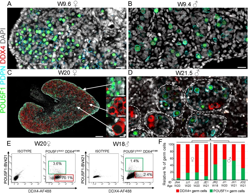
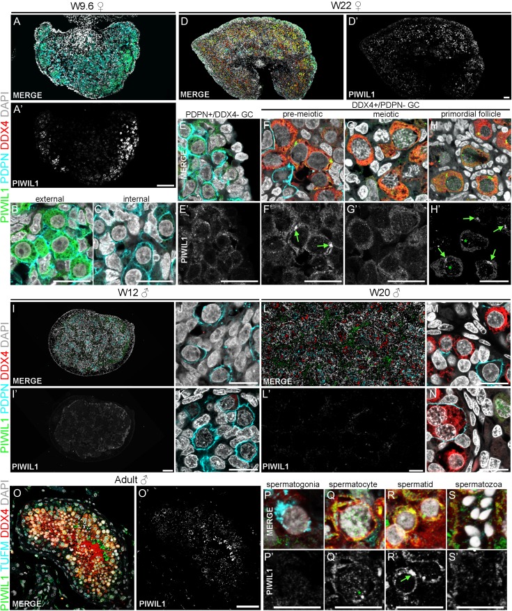
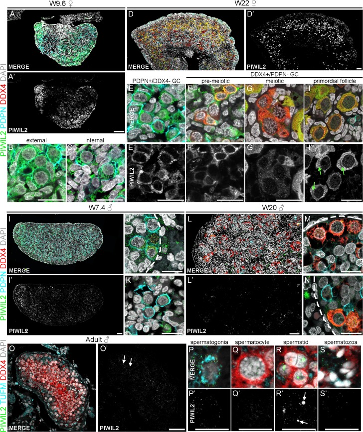
Summary answer: PIWIL1, PIWIL2, PIWIL3 and PIWIL4 were expressed in a sex-specific fashion in human germ cells (GC) during development and adulthood. PIWILs showed a mutually exclusive pattern of subcellular localization. PIWILs were present in the intermitochondrial cement and a single large granule in meiotic GC and their expression was different from that observed in mice, highlighting species-differences.

What is known already: In mice, PIWIL proteins play prominent roles in male infertility. PIWIL mouse mutants show either post-meiotic arrest at the round spermatid stage (PIWIL1) or arrest at the zygotene-pachytene stage of meiosis I (PIWIL2 and PIWIL4) in males, while females remain fertile. Recent studies have reported a robust piRNA pool in human fetal ovary.

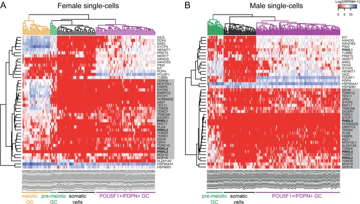
Study design, size, duration: This is a qualitative analysis of PIWILs expression in paraffin-embedded fetal human male (N = 8), female gonads (N = 6) and adult testes (N = 5), and bioinformatics analysis of online available single-cell transcriptomics data of human fetal germ cells (n = 242).

Participants/materials, setting, methods: Human fetal gonads from elective abortion without medical indication and adult testes biopsies were donated for research with informed consent. Samples were fixed, paraffin-embedded and analyzed by immunofluorescence to study the temporal and cellular localization of PIWIL1, PIWIL2, PIWIL3 and PIWIL4.

Main results and the role of chance: PIWIL1, PIWIL2 and PIWIL4 showed a mutually exclusive pattern of subcellular localization, particularly in female oocytes. To our surprise, PIWIL1 immunostaining revealed the presence of a single dense paranuclear body, resembling the chromatoid body of haploid spermatocytes, in meiotic oocytes. Moreover, in contrast to mice, PIWIL4, but not PIWIL2, localized to the intermitochondrial cement. PIWIL3 was not expressed in GC during development. The upregulation of PIWIL transcripts correlated with the transcription of markers associated with piRNAs biogenesis like the TDRDs and HENMT1 in fetal GC.

Large scale data: Non-applicable.

Limitations, reasons for caution: This study is limited by the restricted number of samples and consequently stages analyzed.

Wider implications of the findings: In the germline, PIWILs ensure the integrity of the human genome protecting it from 'parasitic sequences'. This study offers novel insights on the expression dynamics of PIWILs during the window of epigenetic remodeling and meiosis, and highlights important differences between humans and mice, which may prove particularly important to understand causes of infertility and improve both diagnosis and treatment in humans.

Study funding/competing interest(s): M.G.F. was funded by Fundação para a Ciência e Tecnologia (FCT) [SFRH/BD/78689/2011]; N.H. by China Scholarship Council (CSC) [No. 201307040026] and F.W. by Medical Personnel Training Abroad Project of Henan Province [No. 2015022] and S.M.C.d.S.L. by the Netherlands Organization of Scientific Research (NWO) [ASPASIA 015.007.037] and the Interuniversity Attraction Poles-Phase VII [IUAP/PAI P7/14]. The authors have no conflicts of interest to declare.

Keywords: PIWIL; chromatoid body; development; gametogenesis; human; intermitochondrial cement; meiosis; oocyte; spermatogenesis; subcellular localization.


Figures

Figure 1
Figure 1
Expression of PDPN, POU5F1 and DDX4 in human gonads. (A–B) Female gonad (W9.6) (A) and male gonad (W9.4) (B) showing a homogeneous population of double positive POU5F1 and PDPN GC. (C) Female gonad (W20) showing in addition to POU5F1+/PDPN+ GC, a distinct population of DDX4+/POU5F1- GC (pre-meiotic, meiotic and those encapsulated in primordial follicles). (D) Male gonad (W21.5) showing distinct populations of POU5F1+/PDPN+ GC and DDX4+pre-meiotic GC in the seminiferous tubules (dashed line). (E) FACS dot-plots showing female and male gonads immunostained for POU5F1 and DDX4, as well the isotype controls. Gatings show the percentage of POU5F1-positive (green) and DDX4-positive (red) GC. (F) Quantification of relative percentage (%) of POU5F1-positive (green) and DDX4-positive GC per gonad. Identification (ID) and age (weeks) are provided for each gonad. Both the % POU5F1-positive and DDX4-positive GC were statistically different between males and females (*P < 0.05). Scale bars are 20 μm in A, B, D and inserts in C; and 200 μm C.
Figure 2
Figure 2
PIWIL1 expression in the female and male germ cells. (A–N) Histological sections immunostained for PIWIL1 (green), PDPN (cyan) and DDX4 (red) and counterstained with DAPI (white) of female gonad (W9.6) (A–C), female gonad (W22) (D–H), male gonad (W12) (I–K) and male gonad (W20) (L–N). (O–S) Histological sections immunostained for PIWIL1 (green), TUFM (cyan) and DDX4 (red) and counterstained with DAPI (white) of adult human testis. Green arrows point to the single dense paranuclear granule; green asterisks mark nuclear foci of PIWIL1. Scale bars are 500 μm in D, D’; 100 μm A, A’, I, I’, L, L’; 50 μm in H, H’, O, O’; 20 μm in B, C, E–G, E’–G’, J, K, M, N and 10 μm in P–S and P’–S’.
Figure 3
Figure 3
PIWIL4 expression in the female and male germ cells. (A–N) Histological sections immunostained for PIWIL4 (green), PDPN (cyan) and DDX4 (red) and counterstained with DAPI (white) of female gonad (W9.6) (A–C), female gonad (W22) (D–H), male gonad (W7.4) (I–K) and male gonad (W20) (L–N). (O–S) Histological sections immunostained for PIWIL4 (green), TUFM (cyan) and DDX4 (red) and counterstained with DAPI (white) of adult human testis. Green arrows point to intermitochondrial cement. Scale bars are 500 μm in D, D’; 100 μm A, A’, I, I’, L, L’; 50 μm in H, H’, O, O’; 20 μm in B, C, E–G, E’–G’, J, K, M, N and 10 μm in P–S and P’–S’.
Figure 4
Figure 4
Localization of PIWILs relative to the Golgi apparatus and mitochondria. (A–B) Histological sections of human female (W22) (A) and male (W20) (B) gonads immunostained for the Golgi-marker GM130 (green), PIWIL4 (green) or PIWIL2 (green) in combination with DDX4 (cyan), mitochondria-marker TUFM (red) and counterstained with DAPI (white). Yellow arrows point to complexes of Golgi apparatus; yellow dashed areas in oocytes mark the concentration of TUFM+ mitochondria (intermitochondrial cement). Scale bars are 20 μm.
Figure 5
Figure 5
PIWIL2 expression in the female and male germ cells. (A–N) Histological sections immunostained for PIWIL2 (green), PDPN (cyan) and DDX4 (red) and counterstained with DAPI (white) of female gonad (W9.6) (A–C), female gonad (W22) (D–H), male gonad (W7.4) (I–K) and male gonad (W20) (L–N). (O–S) Histological sections immunostained for PIWIL2 (green), TUFM (cyan) and DDX4 (red) and counterstained with DAPI (white) of adult human testis. Green arrows point to intermitochondrial cement; white arrows point to nuclear foci of PIWIL2. Scale bars are 500 μm in D, D’; 100 μm A, A’, I, I’, L, L’; 50 μm in H, H’, O, O’; 20 μm in B, C, E–G, E’–G’, J, K, M, N and 10 μm in P–S and P’–S’.
Figure 6
Figure 6
Unsupervised hierarchical clustering of single-cell human female and male germ cells. (A–B) Heat maps depict the expression of 43 selected germline-specific genes and PIWI/piRNA associated genes (cluster in gray box) in female (n = 93 germ cells and n = 38 somatic cells) and male (n = 149 germ cells and n = 48 somatic cells) single cells from nine different donors from first and second trimester (Guo et al., 2015). Note that the single cells cluster into somatic (black), POU5F1+/PDPN+ germ cells (GC) (violet), pre-meiotic GC (green) and meiotic GC (orange). This dataset contained no oocytes in primordial follicles.

References

    1. Anderson RA, Fulton N, Cowan G, Coutts S, Saunders PT. Conserved and divergent patterns of expression of DAZL, VASA and OCT4 in the germ cells of the human fetal ovary and testis. BMC Dev Biol 2007;7:136. - PMC - PubMed
    1. Aravin AA, Sachidanandam R, Bourc’his D, Schaefer C, Pezic D, Toth KF, Bestor T, Hannon GJ. A piRNA pathway primed by individual transposons is linked to de novo DNA methylation in mice. Mol Cell 2008;31:785–799. - PMC - PubMed
    1. Aravin AA, van der Heijden GW, Castaneda J, Vagin VV, Hannon GJ, Bortvin A. Cytoplasmic compartmentalization of the fetal piRNA pathway in mice. PLoS Genet 2009;5:e1000764. - PMC - PubMed
    1. Bertocchini F, Chuva de Sousa Lopes SM. Germline development in amniotes: a paradigm shift in primordial germ cell specification. BioEssays 2016;38:791–800. - PMC - PubMed
    1. Carmell MA, Girard A, van de Kant HJ, Bourc’his D, Bestor TH, de Rooij DG, Hannon GJ. MIWI2 is essential for spermatogenesis and repression of transposons in the mouse male germline. Dev Cell 2007;12:503–514. - PubMed

Publication types