. 2018;10(2):94-105.
doi: 10.1159/000484257. Epub 2017 Dec 14.

Kallikrein Cleaves C3 and Activates Complement

Affiliations

Kallikrein Cleaves C3 and Activates Complement

Sarah Irmscher et al. J Innate Immun. 2018.

Abstract

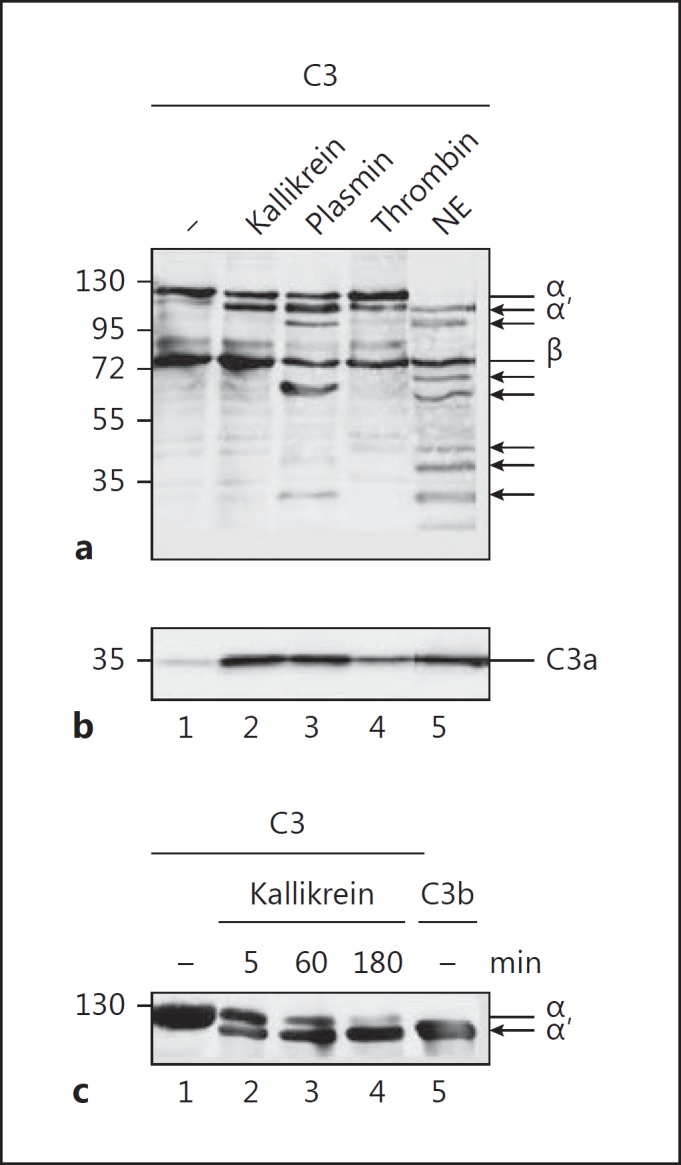
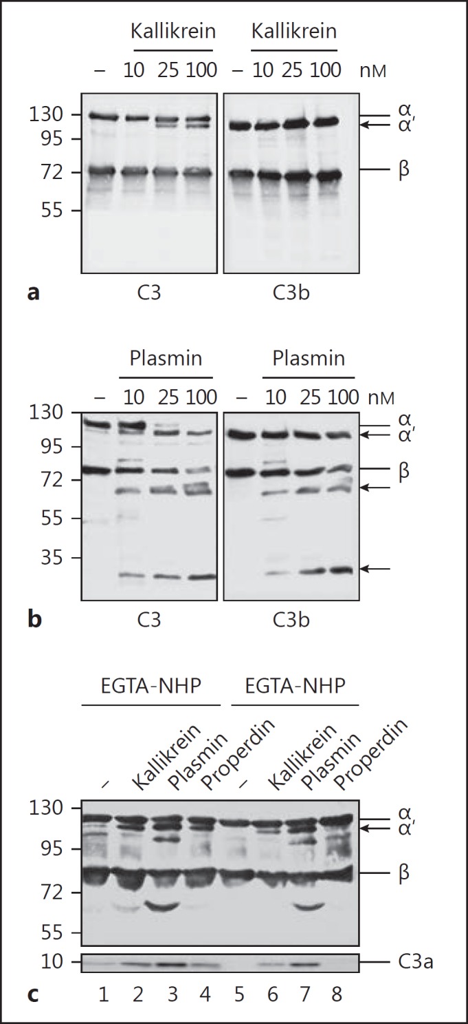
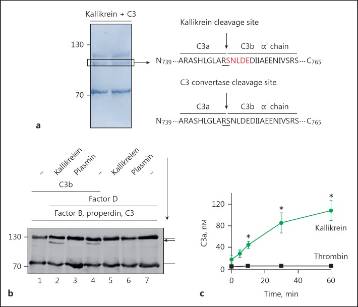
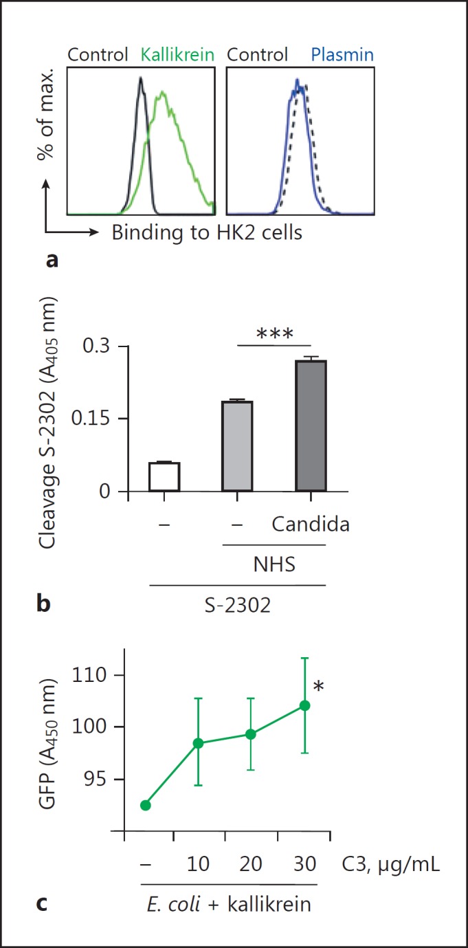
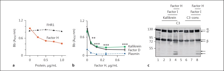
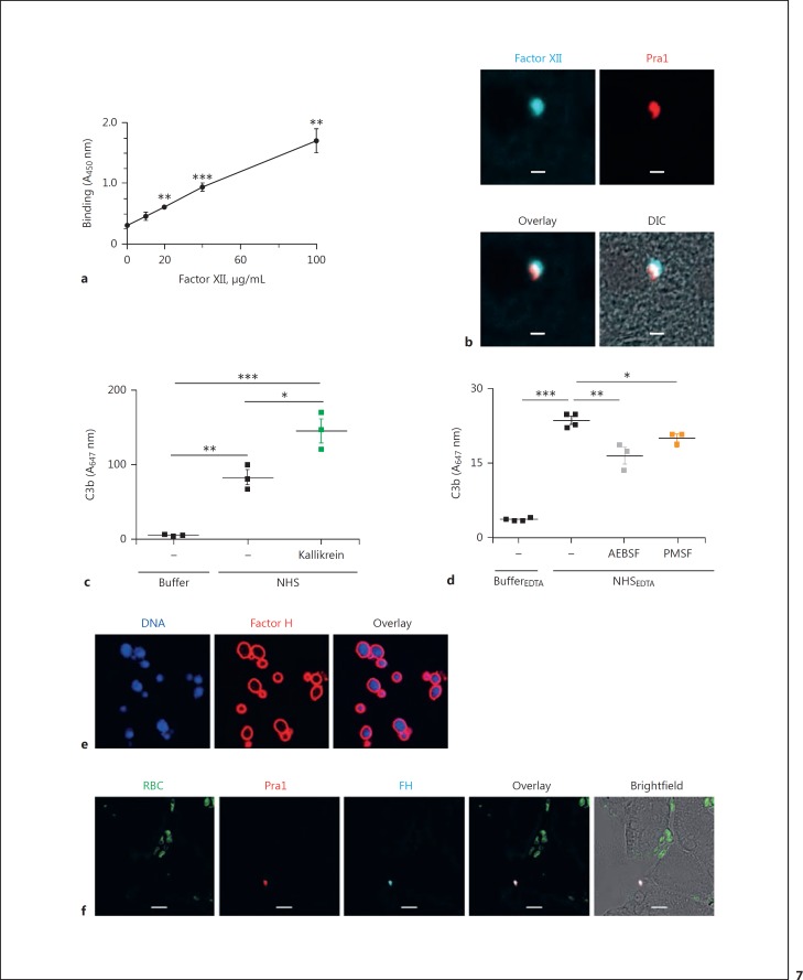
The human plasma contact system is an immune surveillance system activated by the negatively charged surfaces of bacteria and fungi and includes the kallikrein-kinin, the coagulation, and the fibrinolytic systems. Previous work shows that the contact system also activates complement, and that plasma enzymes like kallikrein, plasmin, thrombin, and FXII are involved in the activation process. Here, we show for the first time that kallikrein cleaves the central complement component C3 directly to yield active components C3b and C3a. The cleavage site within C3 is identical to that recognized by the C3 convertase. Also, kallikrein-generated C3b forms C3 convertases, which trigger the C3 amplification loop. Since kallikrein also cleaves factor B to yield Bb and Ba, kallikrein alone can trigger complement activation. Kallikrein-generated C3 convertases are inhibited by factor H; thus, the kallikrein activation pathway merges with the amplification loop of the alternative pathway. Taken together, these data suggest that activation of the contact system locally enhances complement activation on cell surfaces. The human pathogenic microbe Candida albicans activates the contact system in normal human serum. However, C. albicans immediately recruits factor H to the surface, thereby evading the alternative and likely kallikrein-mediated complement pathways.

Keywords: Candida albicans; Complement system; Contact activation; Factor H; Host defense; Kallikrein.


Figures

Fig. 1
Fig. 1
C3 cleavage by proteases. a C3 is cleaved by kallikrein, plasmin, thrombin, and neutrophil elastase (NE). All proteases generate C3b-like (upper panel α′ chain) and C3a-like molecules (b). Incubation of C3 with plasmin (lane 3) and NE (lane 5) generates further cleavage products of C3 as indicated by additional C3 bands in a (arrows) using Western blot analysis. c Kallikrein (100 nM) incubated with C3 (50 nM) for 180 min completely cleaves C3 to C3b-like fragments (lanes 1–4). Purified C3b is shown in lane 5. Proteins were separated by SDS-PAGE and immunoblotted using C3 (a, c) and C3a (b) antibodies. Representative Western blots are shown.
Fig. 2
Fig. 2
Kallikrein generates C3b-like fragments and plasmin degrades C3 and C3b. a Kallikrein (10, 25, and 100 nM) incubated with C3 (50 nM) for 60 min cleaves C3 to C3b-like fragments (left panel) but has no effect on C3b (right panel), as shown by Western blot analysis. b Plasmin (10, 25, and 100 nM) degrades C3 (left panel) and C3b (right panel), and cleavage products of 60- and 30-kDa appear (arrows). c Kallikrein cleaves C3 in EGTA- (inhibitor of the CP) and EDTA- (inhibitor of AP, CP, and LP) buffered human plasma (1%), and C3a-like fragments are generated (lanes 2 and 6), as shown by Western blot analysis. Also, the addition of properdin (350 nM) to EGTA-treated plasma generates active convertases (lane 4), but the addition of properdin to EDTA-treated plasma (lane 8) does not initiate C3 convertases. In contrast to kallikrein, plasmin cleaves C3 to C3b- and C3a-like fragments, but degrades C3 to further fragments of about 100 and 60 kDa (lanes 3 and 7). Representative immune blots from 3 independent experiments are shown (a–c).
Fig. 3
Fig. 3
Kallikrein cleaves C3 like the C3 convertase. a N-terminal sequencing of the kallikrein-generated C3b-like cleavage product (left panel) revealed cleavage of C3 between the amino acids arginine (R) and serine (S), as indicated by the arrow (right panel). The cleavage site by the C3 convertase is indicated. Cleavage of C3 by kallikrein is shown by Coomassie blue staining (left panel). Identified amino acids are boxed. b Immobilized C3b preincubated with factor B and factor D forms a convertase that cleaves C3 to C3b (lane 4) and C3a. Coated C3b preincubated with kallikrein still forms C3 convertases (lane 2), as seen by the appearance of the C3bα′ chain. No C3 convertases formed when coated C3b was preincubated with plasmin (lane 3). The immune blot is representative for 3 independent experiments. The arrow indicates the sequence of incubation. c Kallikrein cleaves C3 more efficiently than similar amounts of thrombin. C3 cleavage was followed over time by the generation of C3a using Western blot, followed by densitometric analysis. Data represent mean values ± SE of 3 independent experiments. One-tailed t test, * p < 0.05.
Fig. 4
Fig. 4
Kallikrein generated C3b deposits on cells. a C3b generated from C3 by kallikrein deposits on HK2 cells. In contrast, plasmin degrades C3. A representative flow cytometry result of 3 independent experiments is shown. bC. albicans activates the contact system. C. albicans was incubated in NHS and kallikrein formation was followed by cleavage of the chromogenic substrate Chromogenix S-2302 by kallikrein. c C3a generated by kallikrein displays antimicrobial action and kills E. coli. GFP-labelled E. coli cells were incubated with C3 and kallikrein, and GFP released by dead E. coli was determined by absorbance. Data in b and c represent mean values ± SE of 3 independent experiments. One-tailed t test, * p < 0.05, *** p < 0.001.
Fig. 5
Fig. 5
Kallikrein cleaves factor B like factor D. a Kallikrein incubated with factor B and C3b (5, 25, 50, or 100 nM) in the presence of NiCl2 reveals the cleavage of factor B to fragments Bb and Ba (lanes 2–5), as demonstrated by immune blotting. Similarly, factor D incubated with C3b and factor B cleaves factor B to Bb and Ba. Factor B cleavage increases with C3b concentrations (lanes 3–5). b Incubation of plasmin with C3b and factor B results in the degradation of factor B (lanes 2–5) and additional bands appear (arrows). c Kallikrein cleaves factor B in a dose-dependent manner, as determined by ELISA, in contrast to plasmin. Data represent mean values ± SD of 3 independent experiments. One-tailed t test, ** p < 0.01, *** p < 0.001. d Kallikrein forms active C3 convertases upon incubation with C3 and factor B. Kallikrein was incubated with C3 and factor B in the presence of NiCl2. Convertase formation was followed by the generation of C3b (lanes 3 and 4, arrow) and in parallel C3a using immunoblotting (e; lanes 3 and 4). Assembly of a C3 convertase by C3b, factor B, factor D, and C3 also shows C3a formation (lane 2).
Fig. 6
Fig. 6
Factor H regulates kallikrein-formed convertases. a Factor H but not FHR1 dissociates kallikrein-formed active C3 convertases. Kallikrein-formed convertases were incubated with increasing amounts of factor H or FHR1, and factor Bb binding was determined by ELISA. One representative experiment out of 3 is shown. b Factor H dissociates kallikrein- and factor D-generated convertases. Plasmin fails to create convertases. Data represent mean values ± SD of 3 independent experiments. One-tailed t test, ** p < 0.01, *** p < 0.001. c Factor H and factor I form iC3b from kallikrein-formed C3b molecules (lane 4). IC3b generation from C3 convertase-formed C3b is shown in lane 8. A representative Western blot of 3 independent experiments is shown.
Fig. 7
Fig. 7
C. albicans activates the contact system and recruits factor H. a Purified factor XII binds to C. albicans in a dose-dependent manner. Data represent mean values ± SE of 3 independent experiments. One-tailed t test, **p < 0.01, *** p < 0.001. b FXII is also recruited to the surface of C. albicans in liver tissue sections derived from C. albicans-infected mice (BALB/c). Blue, FXII; red, fungal protein Pra1. cC. albicans activates complement upon incubation in NHS and C3b is deposited on the surface. The addition of kallikrein enhances C3b deposition. d Inhibition of serine proteases in complement-inactive NHS reduces C3b deposition on C. albicans cells. C. albicans was incubated in complement-inhibited NHS(NHSEDTA) with or without the serine protease inhibitors AEBSF or PMSF. C3b deposition on C. albicans cells was measured by flow cytometry. The data represent means ± SEM of 3–4 independent experiments. * p < 0.05, ** p < 0.01, *** p < 0.001. e Purified factor H binds to the surface of C. albicans cells. Red, factor H; blue, DNA (DAPI). f Factor H is recruited to C. albicans in liver tissues. Green, erythrocytes; red, fungal protein Pra1; blue, mouse factor H. Images were taken with an LSM710 microscope (Zeiss) fitted with a ×40 1.4 NA oil-immersion lens. Scale bars, 10 µm.

Similar articles

Cited by

References

    1. Nickel KF, Renné T. Crosstalk of the plasma contact system with bacteria. Thromb Res. 2012;130:S78–S83. - PubMed
    1. Maas C, Oschatz C, Renné T. The plasma contact system 2.0. Semin Thromb Hemost. 2011;37:375–381. - PubMed
    1. Zerleder S. C1-inhibitor: more than a serine protease inhibitor. Semin Thromb Hemost. 2011;37:362–374. - PubMed
    1. Derkx FH, Bouma BN, Schalekamp MP, Schalekamp MA. An intrinsic factor XII- prekallikrein-dependent pathway activates the human plasma renin-angiotensin system. Nature. 1979;280:315–316. - PubMed
    1. Ekdahl KN, Teramura Y, Hamad OA, Asif S, Duehrkop C, Fromell K, Gustafson E, Hong J, Kozarcanin H, Magnusson PU, Huber-Lang M, Garred P, Nilsson B. Dangerous liaisons: complement, coagulation, and kallikrein/kinin cross-talk act as a linchpin in the events leading to thromboinflammation. Immunol Rev. 2016;274:245–269. - PubMed

Publication types

MeSH terms