Review
. 2018 Feb;30(2):e12565.
doi: 10.1111/jne.12565.

Sex steroid hormones and brain function: PET imaging as a tool for research

Affiliations
Review

Sex steroid hormones and brain function: PET imaging as a tool for research

R Moraga-Amaro et al. J Neuroendocrinol. 2018 Feb.

Abstract

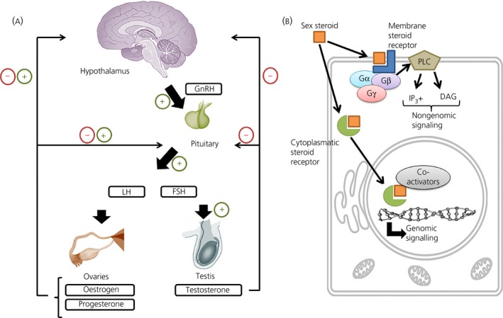
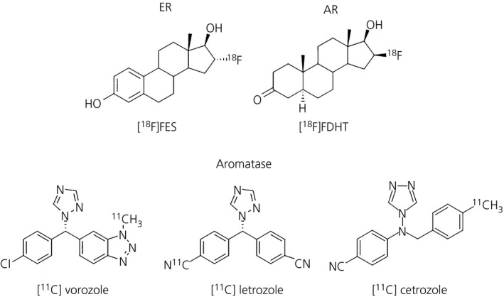
Sex steroid hormones are major regulators of sexual characteristic among species. These hormones, however, are also produced in the brain. Steroidal hormone-mediated signalling via the corresponding hormone receptors can influence brain function at the cellular level and thus affect behaviour and higher brain functions. Altered steroid hormone signalling has been associated with psychiatric disorders, such as anxiety and depression. Neurosteroids are also considered to have a neuroprotective effect in neurodegenerative diseases. So far, the role of steroid hormone receptors in physiological and pathological conditions has mainly been investigated post mortem on animal or human brain tissues. To study the dynamic interplay between sex steroids, their receptors, brain function and behaviour in psychiatric and neurological disorders in a longitudinal manner, however, non-invasive techniques are needed. Positron emission tomography (PET) is a non-invasive imaging tool that is used to quantitatively investigate a variety of physiological and biochemical parameters in vivo. PET uses radiotracers aimed at a specific target (eg, receptor, enzyme, transporter) to visualise the processes of interest. In this review, we discuss the current status of the use of PET imaging for studying sex steroid hormones in the brain. So far, PET has mainly been investigated as a tool to measure (changes in) sex hormone receptor expression in the brain, to measure a key enzyme in the steroid synthesis pathway (aromatase) and to evaluate the effects of hormonal treatment by imaging specific downstream processes in the brain. Although validated radiotracers for a number of targets are still warranted, PET can already be a useful technique for steroid hormone research and facilitate the translation of interesting findings in animal studies to clinical trials in patients.

Keywords: androgen receptor; neuroimaging; oestrogen receptor; positron emission tomography; sex steroid hormones.


Figures

Figure 1
Figure 1
Effects of sex steroids at both physiological and cellular levels. (A) The regulatory processes for the synthesis of sex steroids by the hypothalamic‐pituitary‐gonadal (HPG) axis. The hypothalamus regulates the production of luteinising hormone (LH) and follicle‐stimulating hormone (FSH) via the release of gonadotrophin‐releasing hormone (GnRH). Both LH and FSH stimulate the synthesis and release of oestrogens and progesterone from the ovaries in females, as well as testosterone from the testis in males. At the same time, these sex steroids can regulate the release of GnRH from the hypothalamus, as well as LH and FSH from the pituitary. (B) General scheme of sex steroid effects at cellular level. Sex hormones can bind to either cytoplasmatic receptors or membrane‐associated receptors. When the molecules bind to membrane receptors, the receptor (coupled to G protein subunits complex: Gα, Gβ and Gγ) activates phospholipase C (PLC) to exert a rapid nongenomic responses via the second messengers inositol phosphate 3 (IP 3+) and diacylglycerol (DAG). On the other hand, when they bind to cytoplasmatic receptors, the complex is translocated to the nucleus (with the help of different co‐activators) to exert genomic effects
Figure 2
Figure 2
(A) Positron emission tomography (PET) images of the distribution of the oestrogen receptor tracer 16α‐[18F] fluoro‐17ß‐oestradiol ([18F]FES) in the brain of a healthy postmenopausal woman. The images were acquired 60‐90 min after injection of 200 MBq of [18F]FES. Tracer uptake is presented as kBq/cc images. The pituitary is clearly visible as a hotspot (red) in the sagittal image (top right). In addition, the images mainly show uptake in white matter. (B) PET scan of a naïve female rat brain, 60‐90 min after injection of 25 MBq of [18F]FES. The PET scan is co‐registered with a magnetic resonance imaging template of the brain to provide an anatomical reference. The highlighted spot represents the activity of the tracer in the pituitary/hypothalamus
Figure 3
Figure 3
Chemical structure of tested radiotracers in the brain for both sex hormone receptors and oestrogen synthesis. 16α‐[18F] fluoro‐17ß‐oestradiol ([18F]FES) is the radioligand used for oestrogen receptors (ER). 16ß‐[18F]fluorodihydrotestosterone ([18F]FDHT) corresponds to the tracer tested in the rat brain to visualise androgen receptors (AR). [11C]vorozole, [11C]letrozole and [11C]cetrozole are all tracers for aromatase quantification, which is the enzyme responsible for oestrogen production using testosterone as substrate
Figure 4
Figure 4
(A) 2‐Deoxy‐2‐[18F]fluoro‐d‐glucose ([18F]FDG) positron emission tomography (PET) images of the brain of a healthy woman. The subject had fasted for 6 hours prior to the scan. Static PET images were acquired 30‐35 min after injection of 200 MBq of [18F]FDG. [18F]FDG uptake is presented as standardised uptake values (SUV) and is a surrogate marker for brain glucose metabolism. (B) [18F]FDG PET images of the brain of a healthy female Wistar rat. The images were reconstructed from a 30‐min static scan that was acquired 45 min after i.v. injection of 17 MBq of [18F]FDG. [18F]FDG uptake is presented as the SUV

Similar articles

Cited by

References

    1. Simerly RB. Wired on hormones: endocrine regulation of hypothalamic development. Curr Opin Neurobiol. 2005;15:81‐85. - PubMed
    1. Frick KM, Kim J, Tuscher JJ, Fortress AM. Sex steroid hormones matter for learning and memory: estrogenic regulation of hippocampal function in male and female rodents. Learn Mem. 2015;22:472‐493. - PMC - PubMed
    1. Maeng LY, Milad MR. Sex differences in anxiety disorders: interactions between fear, stress, and gonadal hormones. Horm Behav. 2015;76:106‐117. - PMC - PubMed
    1. Diamanti‐Kandarakis E, Dattilo M, Macut D, et al. Mechanisms in endocrinology : aging and anti‐aging: a combo‐endocrinology overview. Eur J Endocrinol 2017;176:R283‐R308. - PubMed
    1. Handa RJ, Weiser MJ. Gonadal steroid hormones and the hypothalamo‐pituitary‐adrenal axis. Front Neuroendocrinol. 2014;35:197‐220. - PMC - PubMed

Substances

LinkOut - more resources