. 2018 Sep 1;29(7):615-636.
doi: 10.1089/ars.2017.7354. Epub 2018 Jan 30.

Redox-Sensing Under Hypochlorite Stress and Infection Conditions by the Rrf2-Family Repressor HypR in Staphylococcus aureus

Affiliations

Redox-Sensing Under Hypochlorite Stress and Infection Conditions by the Rrf2-Family Repressor HypR in Staphylococcus aureus

Vu Van Loi et al. Antioxid Redox Signal. .

Abstract

Aims: Staphylococcus aureus is a major human pathogen and has to cope with reactive oxygen and chlorine species (ROS, RCS) during infections, which requires efficient protection mechanisms to avoid destruction. Here, we have investigated the changes in the RNA-seq transcriptome by the strong oxidant sodium hypochlorite (NaOCl) in S. aureus USA300 to identify novel redox-sensing mechanisms that provide protection under infection conditions.

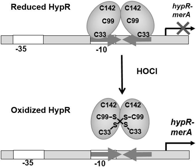
Results: NaOCl stress caused an oxidative stress response in S. aureus as indicated by the induction of the PerR, QsrR, HrcA, and SigmaB regulons in the RNA-seq transcriptome. The hypR-merA (USA300HOU_0588-87) operon was most strongly upregulated under NaOCl stress, which encodes for the Rrf2-family regulator HypR and the pyridine nucleotide disulfide reductase MerA. We have characterized HypR as a novel redox-sensitive repressor that controls MerA expression and directly senses and responds to NaOCl and diamide stress via a thiol-based mechanism in S. aureus. Mutational analysis identified Cys33 and the conserved Cys99 as essential for NaOCl sensing, while Cys99 is also important for repressor activity of HypR in vivo. The redox-sensing mechanism of HypR involves Cys33-Cys99 intersubunit disulfide formation by NaOCl stress both in vitro and in vivo. Moreover, the HypR-controlled flavin disulfide reductase MerA was shown to protect S. aureus against NaOCl stress and increased survival in J774A.1 macrophage infection assays. Conclusion and Innovation: Here, we identified a new member of the widespread Rrf2 family as redox sensor of NaOCl stress in S. aureus that uses a thiol/disulfide switch to regulate defense mechanisms against the oxidative burst under infections in S. aureus. Antioxid. Redox Signal. 29, 615-636.

Keywords: Rrf2; Staphylococcus aureus; hypochlorite stress; redox-sensing regulator.


Conflict of interest statement

No competing financial interests exist.

Figures

<b>FIG. 1.</b>
FIG. 1.
RNA-seq transcriptomics of Staphylococcus aureus USA300 after 30 min of NaOCl stress. For RNA-seq transcriptome profiling, S. aureus USA300 was grown in BMM and treated with 150 μM NaOCl stress for 30 min. The gene expression profile under NaOCl stress is shown as ratio/intensity scatter plot (M/A-plot), which is based on the differential gene expression analysis using DeSeq2. Colored symbols indicate significantly induced (red, magenta, yellow) or repressed (green) transcripts (M-value ≥1.98 or ≤ −1.98; p-value ≤0.05). Black symbols indicate differential transcribed genes below the M-value cutoff of 1.98>M>-1.98 (p ≤ 0.05). Gray symbols denote transcripts with no fold changes after NaOCl stress (p > 0.05). The SaeRS, HypR, MgrA, ArgR, CodY, and QsrR regulons are most strongly upregulated under NaOCl stress. The transcriptome analysis was performed from three biological replicates. The RNA-seq expression data of all genes after NaOCl stress and their regulon classifications are listed in Supplementary Tables S1 and S2. BMM, Belitsky minimal medium; NaOCl, sodium hypochlorite.
<b>FIG. 2.</b>
FIG. 2.
The transcriptome treemap of S. aureus USA300 under NaOCl stress indicates an oxidative stress response and the strong upregulation of the HypR and SaeRS regulons. The transcriptome treemap shows the differential gene expression of S. aureus after exposure to 150 μM NaOCl stress as log2 fold changes (m-values). The genes are classified into operons and regulons based on the RegPrecise database and previous publications (61). Differential gene expression is visualized using a red-blue color code where red indicates log2 fold induction and blue repression of transcription under NaOCl stress. The HypR and SaeRS regulons are most strongly upregulated under NaOCl stress in S. aureus USA300. The induction of the PerR, HrcA, SigmaB, and QsrR regulons reveals an oxidative stress response in S. aureus. The RNA-seq expression data of the selected highly transcribed genes after NaOCl stress and their regulon classifications are listed in Supplementary Table S2.
<b>FIG. 3.</b>
FIG. 3.
The hypR-merA-operon is most strongly upregulated in the RNA-seq transcriptome of S. aureus USA300 under NaOCl stress. The mapped reads for the gene expression profile of the hypR-merA locus under control and NaOCl stress are shown as displayed using the Read-Explorer software. Transcription of the hypR-merA-operon is 180-fold induced under NaOCl stress in S. aureus USA300. The hypR gene encodes for an Rrf2 transcriptional regulator and merA encodes for a pyridine nucleotide disulfide reductase.
<b>FIG. 4.</b>
FIG. 4.
Northern blot analysis of hypR-merA transcription in S. aureus COL under NaOCl, diamide, H2O2, and aldehyde stress and in hypR Cys-Ala mutants. (A) Northern blot analysis was performed using RNA isolated from S. aureus COL wild type before (co) and 15 and 30 min after exposure to 1 mM NaOCl, 2 mM diamide, 10 mM H2O2, 0.75 mM formaldehyde, and 0.5 mM methylglyoxal stress. (B) Transcription of the hypR-merA operon was analyzed in the COL wild-type and in the ΔhypR mutant under 1 mM NaOCl stress indicating strong derepression of hypR-merA transcription under control conditions in the absence of HypR. (C, D) Northern blot analysis of hypR-merA operon transcription in the ΔhypR deletion mutant and in ΔhypR mutants complemented with hypR, hypRC33A, hypRC99A, and hypRC142A before and 15 and 30 min after exposure to 1 mM NaOCl (C) or 2 mM diamide stress (D). The results indicate that Cys33 is required for redox sensing of HypR in vivo. For stress experiments, S. aureus cells were grown in RPMI medium and treated with thiol-reactive compounds at an OD500 of 0.5. The arrows point toward the hypR-merA bicistronic mRNA (1.8 kb) in the wild type or the truncated hypR-merA transcript (TR-merA) (1.5 kb) in the hypR mutant. The methylene blue stain is the RNA loading control indicating the 16S and 23S rRNAs. OD500, optical density at 500 nms. H2O2, hydrogen peroxide.
<b>FIG. 5.</b>
FIG. 5.
Multiple protein sequence alignments of the Rrf2 regulators HypR, SaiR, YwnA, and NsrR (A) and structural modeling of HypR and SaiR in comparison to YwnA and NsrR (B). (A) The protein sequence alignment was performed with ClustalΩ2 and is presented in Jalview. The following protein sequences were aligned and the % identity to HypR is given in parenthesis: HypR (SACOL0641) of S. aureus COL, SaiR (BAS3200) of Bacillus anthracis (20.4%), YwnA (P71036) of Bacillus subtilis (23.48%), and NsrR (Q9L132) of Streptomyces coelicolor (17.86%). Intensity of the blue color gradient is based on 50% sequence identity. The conserved Cys99 in HypR is labeled in red with an asterisk (*). (B) The structural models of HypR and SaiR were generated using SWISS-MODEL (10) and visualized with PyMOL using the template of Bacillus subtilis YwnA (1xd7) that showed 23.5% and 25.78% sequence identity to HypR and SaiR, respectively. For comparison, we show the structures of YwnA (1xd7) and NsrR (5no7) with labels for the conserved Cys97 in YwnA and the 3 FeS cluster coordinating Cys residues (Cys93, Cys99, and Cys105) in NsrR. The FeS cluster of NsrR is displayed in yellow. FeS, iron/sulfur; NsrR, redox sensors for nitric oxide.
<b>FIG. 6.</b>
FIG. 6.
Alignment of the hypR-merA upstream promoter regions with the 12-3-12 bp inverted repeat in Staphylococcus species. The upstream promoter region of the hypR-merA operon includes a 12-3-12 bp inverted repeat sequence that is highly conserved upstream of other hypR-merA homologues across Staphylococci, including S. aureus (SACOL0641), Staphylococcus saprophyticus (SSP2349), Staphylococcus equorum (SE1039_01590), Staphylococcus lugdunensis (SLGD_02231), Staphylococcus haemolyticus (SH2331), and Staphylococcus epidermidis (SE0366). (A) Upper panel shows the reads mapped for the hypR-merA transcript as visualized using ReadExplorer. The putative −10 and −35 promoter sequences are labeled and the 12-3-12 bp conserved inverted repeat is boxed and indicated by arrows. (B) All upstream promoter sequences of hypR-merA homologues were aligned using ClustalΩ2 and presented in Jalview. Intensity of the blue color gradient is based on 50% nucleotide sequence identity. (C) Bottom panel represents the 12-3-12 bp conserved inverted repeat created with WebLogo as HypR operator sequence.
<b>FIG. 7.</b>
FIG. 7.
DNA binding of HypR is inhibited by reversible thiol oxidation under NaOCl and diamide stress in vitro and the effect of HypR Cys mutations on DNA binding and redox sensing. (A) EMSAs were used to analyze the DNA-binding activity of purified HypR, HypRC33A, HypRC99A, and HypRC142A proteins to the hypR-merA upstream promoter region in vitro. Increasing concentrations (0.05–1.25 μM) of HypR were used in the DNA-binding reactions with 0.75 ng of template DNA ranging from −128 to +70 relative to the transcription start site of the hypR-merA operon. (B, C) DNA-binding activity of HypR, HypRC33A, and HypRC142A proteins was inhibited by 1.3–20 μM NaOCl (B) or 10–20 μM diamide (C) and could be restored with 1 mM DTT. This indicates that HypR resembles a redox-sensing regulator that is inactivated due to reversible thiol oxidation. The HypRC99A and HypRC99S mutants were unable to bind to the hypR-merA target promoter. “P” indicates the free probe, “C” is the HypR-DNA complex in the presence of DTT, and “0” indicates the control of HypR-DNA complex after DTT removal before exposure to NaOCl. DTT, dithiothreitol; EMSA, electrophoretic mobility shift assay.
<b>FIG. 8.</b>
FIG. 8.
HypR senses NaOCl stress by intermolecular disulfides, which requires Cys33 and Cys99 in vitro and in vivo. (A, B) The purified HypR wild-type and Cys mutant proteins were treated with increasing NaOCl concentrations in vitro and subjected to nonreducing SDS-PAGE analysis. The reduction of the HypR disulfides after DTT treatment is shown in the reducing SDS-PAGE analysis in Supplementary Figure S3. The HypR intermolecular disulfides (bands 1 and 2) were cut, tryptic digested, and subjected to MALDI-TOF MS/MS analysis as shown in Figure 9 to verify the Cys33-Cys99 disulfide. The bands of the C33A mutant protein that were used for tryptic digestion and MS are boxed and labeled with 3 and 4. The MALDI-TOF results of the C33A mutant tryptic peptides are shown in Supplementary Figure S5B, C. (C, D) For the analysis of HypR disulfides in vivo, we used S. aureus COL with plasmid pRB473-hypR, the ΔhypR deletion mutant and ΔhypR mutant strains complemented with hypR, hypRC33A, hypRC99A, and hypRC142A. S. aureus strains were exposed to NaOCl stress, alkylated with NEM, and protein extracts were subjected to nonreducing Western blot analysis using polyclonal rabbit anti-HypR antibodies. The reducing Western blot analysis of the HypR disulfides and loading controls is shown in Supplementary Figure S6. (E) Nonreducing/reducing diagonal SDS-PAGE and HypR-specific Western blot analysis of alkylated protein extracts were performed to verify the intersubunit disulfides for HypR and the HypRC142A mutant protein under NaOCl stress in vivo, but not in the HypRC33A and HypRC99A mutants. Additional diagonal assays using the HypR immunoprecipitates are shown in Supplementary Figure S7. MALDI-TOF-MS, matrix-assisted laser desorption ionization-time of flight mass spectrometry. NEM, N-ethylmaleimide.
<b>FIG. 9.</b>
FIG. 9.
HypR is oxidized to C33-C99' intermolecular disulfides in vitro as revealed by MALDI-TOF-TOF MS. The intermolecular disulfide band of the oxidized HypR wild-type protein of the SDS-PAGE in Figure 8A (band 1) was tryptic digested. The HypR peptides were measured by MALDI-TOF-TOF MS. (A) The upper panel indicates the MS1 overview scan of all peptides and (B) the lower panel shows the fragmentation of the C33-C99 disulfide peptide (3522.74 Da peak) into the Cys33 (1992.90 Da) and Cys99 peptides (1534.72 Da) in the MS2 scan. The parent ion of the disulfide peptide disappeared in the MS2 scan.
<b>FIG. 10.</b>
FIG. 10.
The flavin disulfide reductase MerA is involved in the defense of S. aureus against hypochlorite stress. Growth phenotype analyses of the S. aureus wild-type (WT), the ΔmerA mutant (A), the merA and merAC43S complemented ΔmerA mutant strains (B) and the hypR mutant (C, D) before and after exposure to sublethal concentrations of 1.5 and 1.75 mM NaOCl stress at an OD500 of 0.5. The NaOCl-sensitive growth phenotype of the merA mutant could be restored by complementation with plasmid-encoded merA and requires Cys43 in the MerA-active site. The results are from four biological replicate experiments. MerA, mercuric ion reductase; OD500, optical density at 500 nm.
<b>FIG. 11.</b>
FIG. 11.
MerA and HypR are both required for NaOCl stress survival in S. aureus. S. aureus COL wild-type (WT), ΔhypR and ΔmerA mutants (A), and their complemented strains and Cys mutants (ΔmerA merA, ΔmerA merAC43S, ΔhypR hypR, ΔhypR hypRC33A) (B, C) were grown in RPMI until an OD500 of 0.5 and treated with 3.5 mM NaOCl. Survival assays were performed by spotting 10 μL of serial dilutions after 3 and 4 h of NaOCl exposure onto LB agar plates. The active site Cys43 of MerA and the redox-sensing Cys33 of HypR are important for NaOCl stress survival. LB, Luria Bertani.
<b>FIG. 12.</b>
FIG. 12.
MerA and HypR are required for survival of S. aureus COL in murine macrophages. The survival of S. aureus strains was analyzed 2, 4, and 24 h postinfection (p.i.) of the murine macrophage cell line J-774A.1 and the CFUs were determined. (A, B) The percentages in survival of the ΔhypR and ΔmerA mutants and complemented strains were calculated in 5–6 biological replicate experiments and the survival at the 2-h time point was set to 100%. (C) The average percentage in survival was calculated for each mutant and complemented strain in relation to the wild type (WT), which was set to 100%. Results of 5–6 biological replicates are presented as scatter dots in (A, B) and mean values of percentage survival in comparison to the wild type (C). Error bars represent the SEM and the statistics was calculated using one-way ANOVA and Tukey's multiple comparisons post hoc test using the GraphPad Prism software. The p-values were determined as follows for the scatter dots (A, B): p = 0.0078 for WT/ΔhypR; p = 0.0303 for WT/ΔmerA; p = 0.0461 for WTpRB473/ΔhypRhypR; and p = 0.1234 for WTpRB473/ΔmerAmerA. For the percentage survival (C), the p-values were determined as p < 0.0001 for WT/ΔhypR, p = 0.0012 for WT/ΔmerA, p = 0.0511 for WTpRB473/ΔhypRhypR, p = 0.4684 for WTpRB473/ΔmerAmerA, p = 0.0459 for ΔhypRhypRhypR, and p = 0.0725 for ΔmerAmerAmerA. Symbols are defined as follows: nsp > 0.05; *p ≤ 0.05; **p ≤ 0.01; and ****p ≤ 0.0001. CFU, colony-forming unit.
<b>FIG. 13.</b>
FIG. 13.
Redox-sensing mechanism of HypR under hypochlorite stress during infection conditions in S. aureus. HypR controls the flavin disulfide reductase MerA, essential for growth and survival under hypochlorite stress and in macrophage infection assays in S. aureus. Cys33 and Cys99 of HypR are required for redox sensing in vivo. Under NaOCl stress, HypR is oxidized to Cys33-Cys99 intersubunit disulfides leading to derepression of hypR-merA transcription.

Similar articles

Cited by

References

    1. Abomoelak B, Hoye EA, Chi J, Marcus SA, Laval F, Bannantine JP, Ward SK, Daffe M, Liu HD, and Talaat AM. mosR, a novel transcriptional regulator of hypoxia and virulence in Mycobacterium tuberculosis. J Bacteriol 191: 5941–5952, 2009 - PMC - PubMed
    1. Antelmann H. and Helmann JD. Thiol-based redox switches and gene regulation. Antioxid Redox Signal 14: 1049–1063, 2011 - PMC - PubMed
    1. Archer GL. Staphylococcus aureus: a well-armed pathogen. Clin Infect Dis 26: 1179–1181, 1998 - PubMed
    1. Argyrou A. and Blanchard JS. Flavoprotein disulfide reductases: advances in chemistry and function. Prog Nucleic Acid Res Mol Biol 78: 89–142, 2004 - PubMed
    1. Arnaud M, Chastanet A, and Debarbouille M. New vector for efficient allelic replacement in naturally nontransformable, low-GC-content, gram-positive bacteria. Appl Environ Microbiol 70: 6887–6891, 2004 - PMC - PubMed

Publication types

MeSH terms

LinkOut - more resources