. 2017 Dec 13;8(1):279.
doi: 10.1186/s13287-017-0729-5.

Transplantation of dental pulp stem cells improves long-term diabetic polyneuropathy together with improvement of nerve morphometrical evaluation

Affiliations

Transplantation of dental pulp stem cells improves long-term diabetic polyneuropathy together with improvement of nerve morphometrical evaluation

Maiko Omi et al. Stem Cell Res Ther. .

Abstract

Background: Although previous reports have revealed the therapeutic potential of stem cell transplantation in diabetic polyneuropathy, the effects of cell transplantation on long-term diabetic polyneuropathy have not been investigated. In this study, we investigated whether the transplantation of dental pulp stem cells (DPSCs) ameliorated long-term diabetic polyneuropathy in streptozotocin (STZ)-induced diabetic rats.

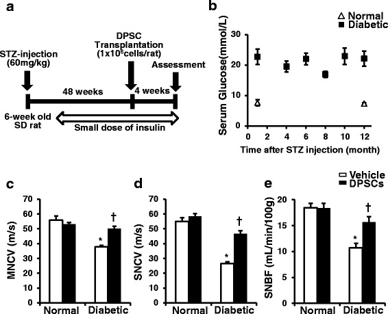
Methods: Forty-eight weeks after STZ injection, we transplanted DPSCs into the unilateral hindlimb skeletal muscles. Four weeks after DPSC transplantation (i.e., 52 weeks after STZ injection) the effects of DPSC transplantation on diabetic polyneuropathy were assessed.

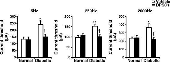
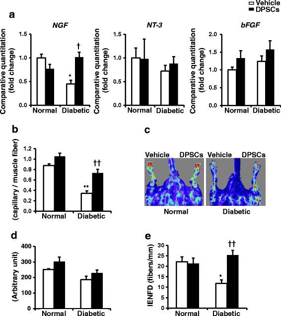
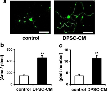
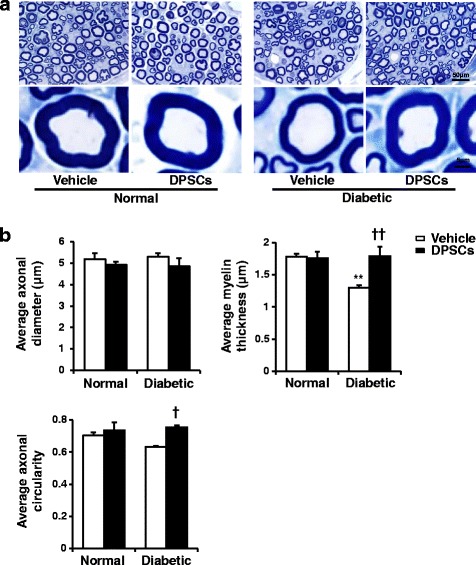
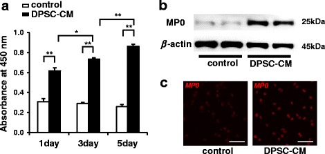
Results: STZ-induced diabetic rats showed significant reductions in the sciatic motor/sensory nerve conduction velocity, increases in the current perception threshold, and decreases in capillary density in skeletal muscles and intra-epidermal nerve fiber density compared with normal rats, all of which were ameliorated by DPSC transplantation. Furthermore, sural nerve morphometrical analysis revealed that the transplantation of DPSCs significantly increased the myelin thickness and area. DPSC-conditioned media promoted the neurite outgrowth of dorsal root ganglion neurons and increased the viability and myelin-related protein expression of Schwann cells.

Conclusions: These results indicated that the transplantation of DPSCs contributed to the neurophysiological and neuropathological recovery from a long duration of diabetic polyneuropathy.

Keywords: Cell transplantation; Dental pulp stem cells; Diabetic polyneuropathy; Nerve repair.


Conflict of interest statement

Ethics approval

All experimental protocols were approved by the Institutional Animal Care and Use Committees of Aichi Gakuin University (AGUD 059) and were conducted in accordance with the United States Public Health Service’s Policy on Humane Care and Use of Laboratory Animals. All efforts were made to minimize animal suffering.

Consent for publication

The authors declare that they consent to publication.

Competing interests

The authors declare that they have no competing interests.

Publisher’s Note

Springer Nature remains neutral with regard to jurisdictional claims in published maps and institutional affiliations.

Figures

Fig. 1
Fig. 1
Characterization and differentiation of DPSCs. a DPSCs derived from 6-week-old Sprague-Dawley rats were positive for mesenchymal stem cell markers (CD29, CD49d, CD90) and negative for hematopoietic markers (CD34 and CD45) (black area). Isotype-identical antibodies served as the controls (white area). b DPSCs could differentiate into adipogenic, osteogenic, and chondrogenic lineages in vitro. For discrimination, Oil Red O and fatty acid-binding protein-4 were used for adipocytes, Alizarin Red and osteocalcin for osteoblasts, and aggrecan for chondrocytes
Fig. 2
Fig. 2
Experimental animal protocol and clinical and neurophysiological measurements. a Dental pulp stem cells (DPSCs) were transplanted into unilateral hind limb skeletal muscles 48 weeks after streptozotocin (STZ) injection, and neurophysiological assessments were performed 4 weeks after DPSC transplantation. Small doses of insulin pellet were administered subcutaneously once a month from 8 weeks after STZ injection to avoid excess hyperglycemia. b Blood glucose concentrations during the experiment. Blood glucose levels were determined at the indicated time points. c Sciatic motor nerve conduction velocity (MNCV). MNCV was measured between the ankle and sciatic notch. d Sciatic sensory nerve conduction velocity (SNCV). SNCV was measured between the ankle and knee; n = 5. e Sciatic nerve blood flow (SNBF). SNBF was measured using a Laser Doppler Blood Flow Meter. The results are means ± SEM. *P < 0.01, vs. vehicle-injected side of normal rats; P < 0.01, vs. vehicle-injected side of diabetic rats. SD Sprague-Dawley
Fig. 3
Fig. 3
The functional integrity of the sensory nerve fibers was assessed by the perception threshold (CPT). The CPT was quantified at each frequency by stimulating Aβ fibers at 2000 Hz, Aδ fibers at 250 Hz, and C fibers at 5 Hz using the Neurometer; n = 5. The results are means ± SEM. *P < 0.05, **P < 0.01, vs. vehicle-injected side of normal rats; P < 0.05, vs. vehicle-injected side of diabetic rats. DPSCs dental pulp stem cells
Fig. 4
Fig. 4
mRNA expressions, capillary density/blood flow in the hind limb and intra-epidermal nerve fiber density. a mRNA expression in the hind limb skeletal muscles. Four weeks after the transplantation of dental pulp stem cells (DPSCs) (the duration of diabetes was 52 weeks), the mRNA expression levels of nerve growth factor (NGF), neurotrophin 3 (NT-3), and basic fibroblast growth factor (bFGF) in the hind limb skeletal muscles were evaluated by real-time quantitative PCR. The results are means ± SEM; n = 7. *P < 0.05, vs. vehicle-injected side of normal rats; P < 0.05, vs. vehicle-injected side of diabetic rats. b The capillary endothelial cells were stained with the anti-von Willebrand factor (vWF) polyclonal antibody. Quantitative analyses for the capillary/muscle fiber ratio of the vehicle-injected and DPSC-transplanted side of skeletal muscles in normal and diabetic rats. The results are means ± SEM. **P < 0.01, vs. vehicle-injected side of normal rats; †† P < 0.01, vs. vehicle-injected side of diabetic rats. c Representative LDPI of the hind limb blood flow of rats. DPSC transplantation increased blood flow (yellow to red color) in the DPSC-injected side of the hind limb of diabetic rats. d Computer-assisted quantitative analyses of hind limb blood flow in normal and diabetic rats. e Intra-epidermal nerve fiber density (IENFD) was evident in both the epidermis and dermis of foot skin by fluorescent imaging for PGP9.5. Intra-epidermal nerve fiber profiles were counted blindly by three independent investigators and the average values were used. The results are means ± SEM. *P < 0.05, vs. vehicle-injected side of normal rats; †† P < 0.01, vs. vehicle-injected side of diabetic rats; n = 7
Fig. 5
Fig. 5
a Dental pulp stem cell-conditioned media (DPSC-CM) promoted neurite outgrowth of DRG neurons. DRG neurons (stained with neurofilament heavy-chain immunofluorescence) extended neurites, and the extensions were significantly promoted in the presence of DPSC-CM compared with that in the absence of DPSC-CM. Scale bars = 100 μm. The total length (b) and joint number (c) of neurite outgrowths in DRG neurons were calculated using a computed image analysis system (Angiogenesis Image Analyzer Ver. 2, KURABO Industries, Osaka, Japan). The results are means ± SEM. **P < 0.01, vs. control group
Fig. 6
Fig. 6
a Representative semi-thin cross-sections of the sural nerves of rats. Toluidine blue stained. b The morphometry of the myelinated nerve fibers. There were no significant differences in the diameter and circularity between the normal and diabetic rats. Myelin thickness was less in the diabetic rats compared with the normal rats. The transplantation of dental pulp stem cells (DPSCs) significantly increased the myelin thickness and circularity. The results are means ± SEM. **P < 0.01, vs. vehicle-injected side of normal rats; P < 0.05, †† P < 0.01, vs. vehicle-injected side of diabetic rats; n = 5
Fig. 7
Fig. 7
Dental pulp stem cell-conditioned media (DPSC-CM) promoted Schwann cell viability and myelin formation. a The effects of DPSC-CM on Schwann cell viability were measured by CCK-8 analysis. The results are means ± SEM. *P < 0.05, **P < 0.01; n = 12. b The protein expression of myelin protein zero (MP0), a related protein for the myelination of Schwann cells, was assessed after a 5-day incubation with DPSC-CM by Western blotting. c Microscopic analysis of MP0-positive myelin segments in Schwann cells treated with DPSC-CM further revealed the expression of MP0 compared with that in the control

References

    1. Greene D. The pathogenesis and prevention of diabetic neuropathy and nephropathy. Metabolism. 1988;37:25–29. doi: 10.1016/0026-0495(88)90184-9. - DOI - PubMed
    1. Sundkvist G, Dahlin LB, Nilsson H, Eriksson KF, Lindgärde F, Rosen I, et al. Sorbitol and myo-inositol levels and morphology of sural nerve in relation to peripheral nerve function and clinical neuropathy in men with diabetic, impaired, and normal glucose tolerance. Diabetic Med. 2000;17:259–268. doi: 10.1046/j.1464-5491.2000.00261.x. - DOI - PubMed
    1. Vincent AM, Callaghan BC, Smith AL, Feldman EL. Diabetic neuropathy: cellular mechanisms as therapeutic targets. Nature Rev Neurol. 2011;7:573–583. doi: 10.1038/nrneurol.2011.137. - DOI - PubMed
    1. Ziegler D. Painful diabetic neuropathy: treatment and future aspects. Diabetes Metab Res Rev. 2008;24(Suppl 1):S52–S57. doi: 10.1002/dmrr.817. - DOI - PubMed
    1. Hosseini A, Abdollahi M. Diabetic neuropathy and oxidative stress: therapeutic perspectives. Oxid Med Cell Longev. 2013;2013:168039. doi: 10.1155/2013/168039. - DOI - PMC - PubMed

MeSH terms