. 2018 Feb 16;293(7):2534-2545.
doi: 10.1074/jbc.M117.815316. Epub 2017 Dec 13.

An allosteric binding site of the α7 nicotinic acetylcholine receptor revealed in a humanized acetylcholine-binding protein

Affiliations

An allosteric binding site of the α7 nicotinic acetylcholine receptor revealed in a humanized acetylcholine-binding protein

Florian Delbart et al. J Biol Chem. .

Abstract

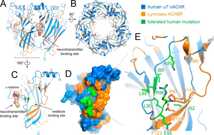
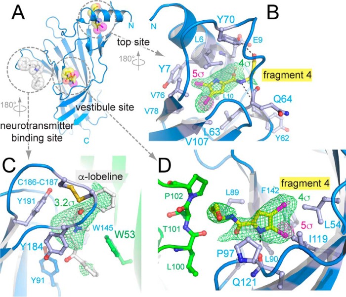
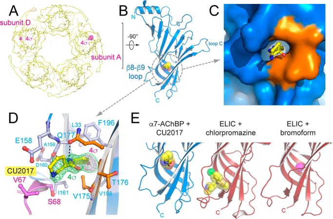
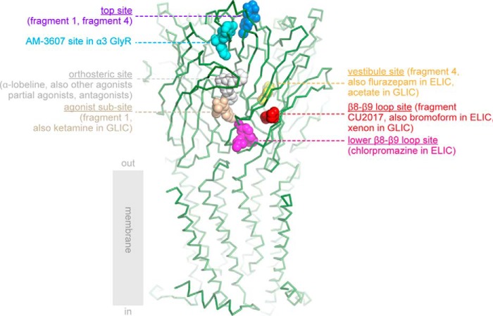
Nicotinic acetylcholine receptors (nAChRs) belong to the family of pentameric ligand-gated ion channels and mediate fast excitatory transmission in the central and peripheral nervous systems. Among the different existing receptor subtypes, the homomeric α7 nAChR has attracted considerable attention because of its possible implication in several neurological and psychiatric disorders, including cognitive decline associated with Alzheimer's disease or schizophrenia. Allosteric modulators of ligand-gated ion channels are of particular interest as therapeutic agents, as they modulate receptor activity without affecting normal fluctuations of synaptic neurotransmitter release. Here, we used X-ray crystallography and surface plasmon resonance spectroscopy of α7-acetylcholine-binding protein (AChBP), a humanized chimera of a snail AChBP, which has 71% sequence similarity with the extracellular ligand-binding domain of the human α7 nAChR, to investigate the structural determinants of allosteric modulation. We extended previous observations that an allosteric site located in the vestibule of the receptor offers an attractive target for receptor modulation. We introduced seven additional humanizing mutations in the vestibule-located binding site of AChBP to improve its suitability as a model for studying allosteric binding. Using a fragment-based screening approach, we uncovered an allosteric binding site located near the β8-β9 loop, which critically contributes to coupling ligand binding to channel opening in human α7 nAChR. This work expands our understanding of the topology of allosteric binding sites in AChBP and, by extrapolation, in the human α7 nAChR as determined by electrophysiology measurements. Our insights pave the way for drug design strategies targeting nAChRs involved in ion channel-mediated disorders.

Keywords: Cys-loop receptor; X-ray crystallography; allosteric modulation; electrophysiology; fragment-based screening; ligand-gated ion channel; neurotransmitter; nicotinic acetylcholine receptors (nAChR); structure-based drug discovery.


Conflict of interest statement

The authors declare that they have no conflicts of interest with the contents of this article

Figures

Figure 1.
Figure 1.
X-ray crystal structure of α7-AChBPVS in complex with α-lobeline. A, schematic representation of the α7-AChBPVS pentamer as seen perpendicular to the 5-fold symmetry axis. Residues belonging to the human α7 nAChR are colored in blue and residues from Lymnaea AChBP are colored orange. The partial agonist α-lobeline is bound to the neurotransmitter (orthosteric)-binding site of the subunit interface facing the viewer and is shown in sphere representation. Carbon atoms are colored in wheat, nitrogen in blue, and oxygen in red. B, view from (A) 90° rotated with the top (N-terminal side) of the pentamer facing the viewer. C and D, schematic and surface representation of a single subunit oriented toward the vestibule-binding site (180° rotated from A), which lies opposite to the neurotransmitter-binding site and is accessible via the receptor vestibule. Additional amino acid residues that were mutated into corresponding residues from the human α7 nAChR are colored in green. E, detailed view of the vestibule-binding site with key side chains shown in stick representation. Orange/blue residues are colored coded as in previous panels and green residues correspond to additional humanizing mutations.
Figure 2.
Figure 2.
X-ray crystal structure of α7-AChBPVS in complex with α-lobeline and allosteric binder fragment 4. A, schematic representation of a single α7-AChBPVS subunit as seen toward the vestibule site. α-Lobeline is bound to the neurotransmitter-binding site and is shown in white transparent sphere and stick representation. Fragment 4 is bound to two different allosteric sites, the vestibule site and the top site and is shown in yellow (carbon), blue (nitrogen), red (oxygen), and magenta (bromine). B, detailed view of the amino acid interactions formed by fragment 4 at the top site. The green mesh is the 5-fold averaged simple difference density at a contour level of 4σ, the magenta mesh is the anomalous difference density at 5σ. C, detailed view of amino acid interactions formed by α-lobeline at the neurotransmitter binding site. The principal subunit is shown in blue, the complementary subunit in green. The highly conserved aromatic side chains are shown as ball and sticks. The green mesh is the 5-fold averaged simple difference density at 3.2σ. D, detailed view of amino acid interactions formed by fragment 4 at the vestibule site. Most interactions are intrasubunit interactions (blue), whereas 3 residues come from a neighboring subunit (green). The green mesh is the 5-fold averaged simple difference density at a contour level of 4σ, the magenta mesh is the anomalous difference density at 5σ.
Figure 3.
Figure 3.
X-ray crystal structure of α7-AChBPVS in complex with allosteric binder fragment CU2017. A, yellow ribbon representation of the α7-AChBPVS pentamer as seen along the 5-fold symmetry axis with the bottom (C-terminal side) of the pentamer pointing toward the viewer. The magenta mesh is the anomalous difference density at a contour level of 4σ. B, schematic representation of the A subunit with fragment CU2017 bound to the β8–β9 loop site. Fragment CU2017 is shown in transparent ball & stick representation in yellow (carbon), blue (nitrogen), red (oxygen), and magenta (bromine). C and D, detailed surface and ball and stick representation of the β8–β9 loop site. Residues belonging to the human α7 nAChR are colored in blue and residues from Lymnaea AChBP are colored in orange. Residues Val-67 and Ser-68 (pink) are from a neighboring pentamer in the crystal packing. In D, the green mesh is the simple difference density at a contour level of 4σ, the magenta mesh is the anomalous difference density at 4σ. E, comparison of the β8–β9 loop site occupied by fragment CU2017 and previously published structures of the Erwinia ligand-gated ion channel ELIC in complex with chlorpromazine (28) (PDB code 5lg3) or bromoform (27) (PDB code 3zkr).
Figure 4.
Figure 4.
Electrophysiological characterization of allosteric fragment CU2017 on human α7 nAChRs expressed in Xenopus oocytes. A, agonist-evoked responses of human α7 nAChRs using 200 μm acetylcholine, first applied alone and then co-applied with increasing concentrations of CU2017 (5–200 μm). B, plot of the peak current responses as a function of fragment concentration and a curve fit of the data with the Hill equation. Data are shown as a scatter plot (red circles) and the mean ± S.D. of 16 independent experiments. The Hill plot has an IC50 value of 26.7 ± 1.6 μm and a coefficient of 1.3 ± 0.1 (n = 16).
Figure 5.
Figure 5.
Overview of ligand-binding sites in α7-AChBP and ligand-binding domains of other Cys-loop receptors. The crystal structure of α7-AChBPVS is shown in green ribbon representation and was superposed onto the extracellular domain of the human α4β2 nAChR structure (13) (PDB code 5kxi). Only the transmembrane domain of the α4β2 receptor is shown for illustrative purposes. The different binding sites are indicated according to their respective bound ligands in different Cys-loop receptor structures and corresponding PDB accession codes are mentioned between parentheses: top site in blue, α7-AChBP + fragment 1 (17) (code 5afj) and α7-AChBPVS + fragment 4 (this study) (code 5oug); AM-3607 site in cyan, α3 GlyR + AM-3607 (43) (code 5tin); orthosteric site in white, example α7-AChBPVS + α-lobeline (code 5ouh); agonist subsite in wheat, α7-AChBP + fragment 1 (17) (code 5afj) and GLIC + ketamine (49) (code 4f8h); β8–β9 loop site in red, α7-AChBPVS + fragment CU2017 (this study) (code 5oui), ELIC + bromoform (27) (code 3zkr) and locally closed GLIC + xenon (47) (code 4zzb); lower part of the β8–β9 loop site in magenta, ELIC + chlorpromazine (28) (code 5lg3); vestibule site in yellow, AChBPVS + fragment 4 structure (this study) (code 5oug), ELIC + flurazepam (18) (code 2yoe), and GLIC + bromoacetate (44) (code 4qh1).

Similar articles

Cited by

References

    1. Thompson A. J., Lester H. A., and Lummis S. C. R. (2010) The structural basis of function in Cys-loop receptors. Quart. Rev. Biophys. 43, 449–499 10.1017/S0033583510000168 - DOI - PubMed
    1. Miller P. S., and Smart T. G. (2010) Binding, activation and modulation of Cys-loop receptors. Trends Pharmacol. Sci. 31, 161–174 10.1016/j.tips.2009.12.005 - DOI - PubMed
    1. Nys M., Kesters D., and Ulens C. (2013) Structural insights into Cys-loop receptor function and ligand recognition. Biochem. Pharmacol. 86, 1042–1053 10.1016/j.bcp.2013.07.001 - DOI - PubMed
    1. Dineley K. T., Pandya A. A., and Yakel J. L. (2015) Nicotinic ACh receptors as therapeutic targets in CNS disorders. Trends Pharmacol. Sci. 36, 96–108 10.1016/j.tips.2014.12.002 - DOI - PMC - PubMed
    1. Plested A. J. (2016) Structural mechanisms of activation and desensitization in neurotransmitter-gated ion channels. Nat. Struct. Mol. Biol. 23, 494–502 10.1038/nsmb.3214 - DOI - PubMed

Publication types

Substances

LinkOut - more resources