

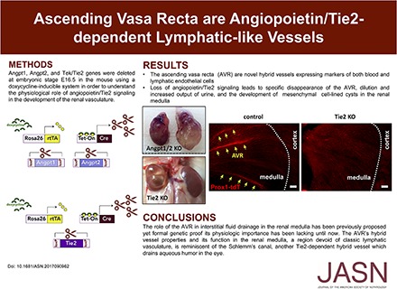
Ascending Vasa Recta Are Angiopoietin/Tie2-Dependent Lymphatic-Like Vessels
- PMID: 29237738
- PMCID: PMC5875961
- DOI: 10.1681/ASN.2017090962
Ascending Vasa Recta Are Angiopoietin/Tie2-Dependent Lymphatic-Like Vessels
Abstract
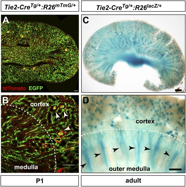
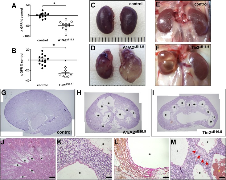
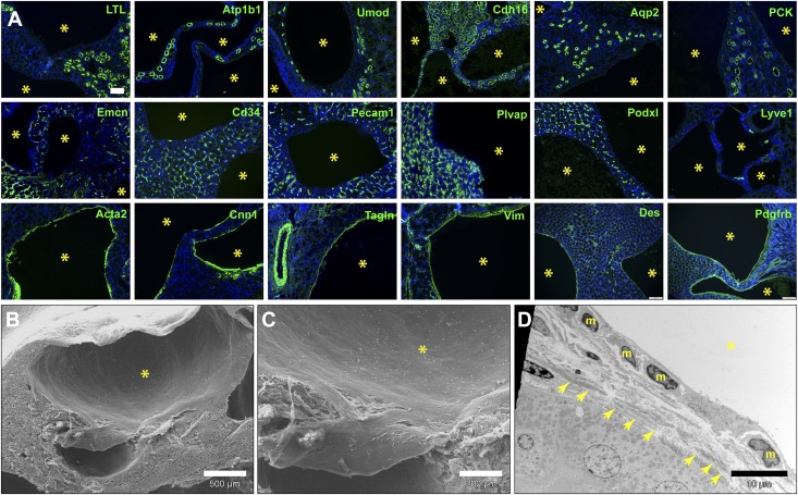
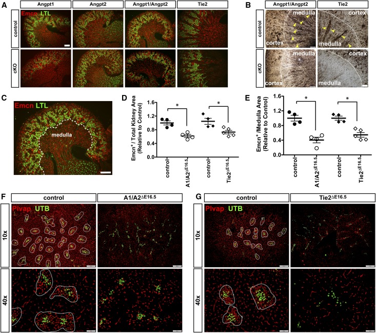
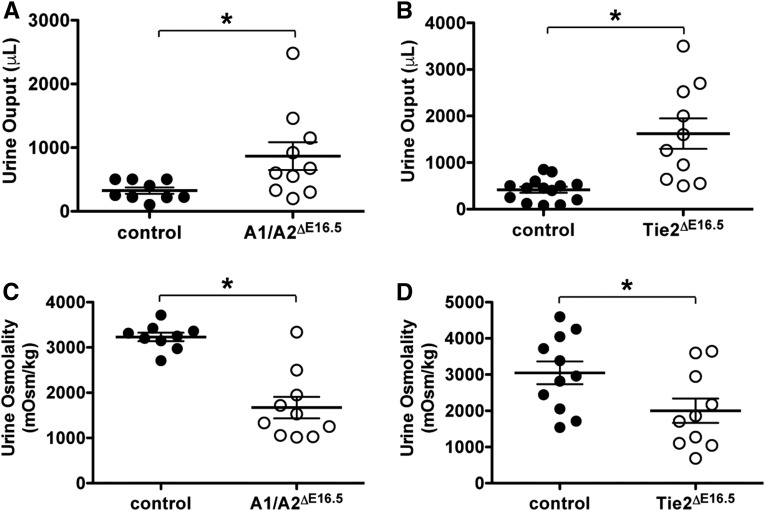
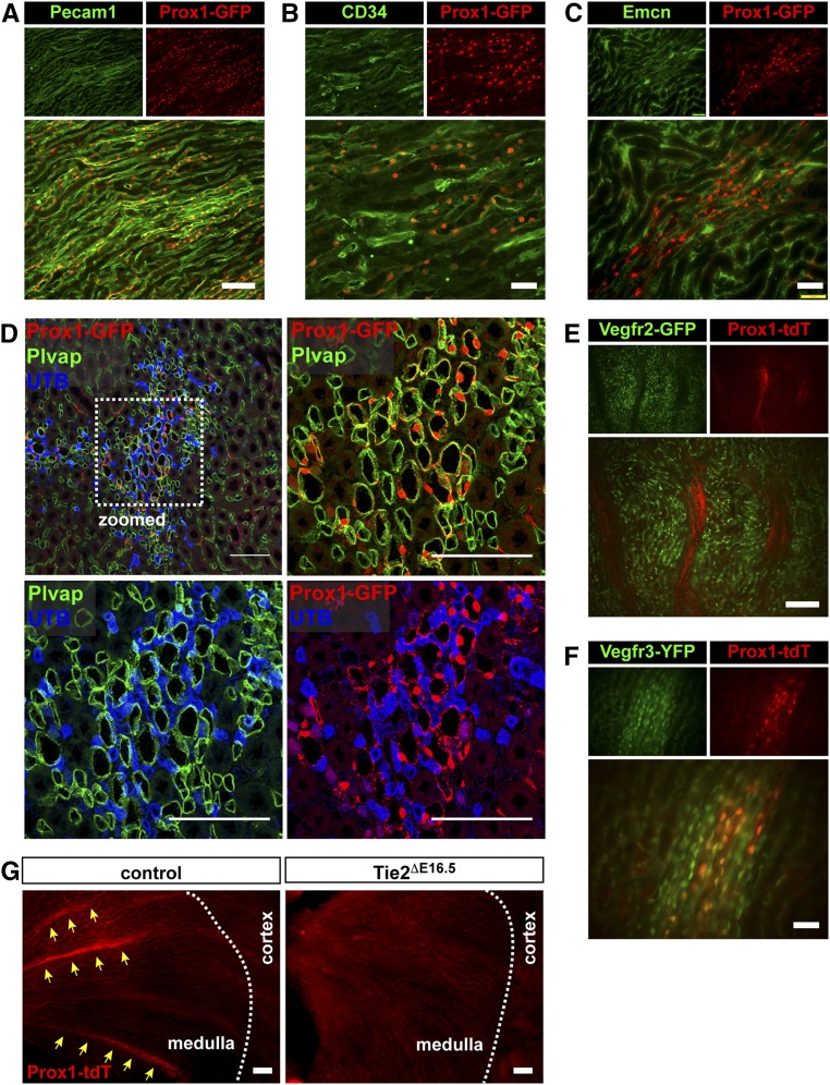
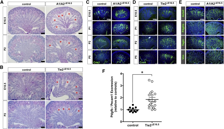
Urinary concentrating ability is central to mammalian water balance and depends on a medullary osmotic gradient generated by a countercurrent multiplication mechanism. Medullary hyperosmolarity is protected from washout by countercurrent exchange and efficient removal of interstitial fluid resorbed from the loop of Henle and collecting ducts. In most tissues, lymphatic vessels drain excess interstitial fluid back to the venous circulation. However, the renal medulla is devoid of classic lymphatics. Studies have suggested that the fenestrated ascending vasa recta (AVRs) drain the interstitial fluid in this location, but this function has not been conclusively shown. We report that late gestational deletion of the angiopoietin receptor endothelial tyrosine kinase 2 (Tie2) or both angiopoietin-1 and angiopoietin-2 prevents AVR formation in mice. The absence of AVR associated with rapid accumulation of fluid and cysts in the medullary interstitium, loss of medullary vascular bundles, and decreased urine concentrating ability. In transgenic reporter mice with normal angiopoietin-Tie2 signaling, medullary AVR exhibited an unusual hybrid endothelial phenotype, expressing lymphatic markers (prospero homeobox protein 1 and vascular endothelial growth factor receptor 3) as well as blood endothelial markers (CD34, endomucin, platelet endothelial cell adhesion molecule 1, and plasmalemmal vesicle-associated protein). Taken together, our data redefine the AVRs as Tie2 signaling-dependent specialized hybrid vessels and provide genetic evidence of the critical role of AVR in the countercurrent exchange mechanism and the structural integrity of the renal medulla.
Keywords: Tie2; angiopoietin; ascending vasa recta; countercurrent exchange; fluid homeostasis; lymphatic.
Copyright © 2018 by the American Society of Nephrology.
Figures
Comment in
-
Unique Gene Expression in Developing Ascending Vasa Recta: A Tale of Tie.J Am Soc Nephrol. 2018 Apr;29(4):1073-1074. doi: 10.1681/ASN.2018020190. Epub 2018 Mar 12. J Am Soc Nephrol. 2018. PMID: 29531096 Free PMC article. No abstract available.
References
-
- Munger KA, Maddox DA, Brenner BM, Kost CK: The renal circulations and glomerular ultrafiltration. In: Brenner & Rector’s the Kidney, 10th Ed., edited by Skorecki K, Chertow GM, Marsden PA, Taal MW, Yu ASL, Philadelphia, Elsevier, 2016, pp 83–111
-
- Pallone TL, Chunhua C: Renal cortical and medullary microcirculations: Structure and function. In: Seldin and Giebisch’s the Kidney, 5th Ed., edited by Alpern RJ, Moe OW, Caplan MJ, London, Academic Press, 2013, pp 803–857
-
- Kriz W, Kaissling B: Structural organization of the mammalian kidney. In: Seldin and Giebisch’s the Kidney, 5th Ed., edited by Alpern RJ, Moe OW, Caplan MJ, Amsterdam, Elsevier, 2013, pp 595–691
-
- Schwartz MM, Karnovsky MJ, Vehkatachalam MA: Ultrastructural differences between rat inner medullary descending and ascending vasa recta. Lab Invest 35: 161–170, 1976 - PubMed
-
- Pallone TL, Turner MR, Edwards A, Jamison RL: Countercurrent exchange in the renal medulla. Am J Physiol Regul Integr Comp Physiol 284: R1153–R1175, 2003 - PubMed
Publication types
MeSH terms
Substances
Grants and funding
LinkOut - more resources
Full Text Sources
Other Literature Sources
Molecular Biology Databases
Research Materials
Miscellaneous
