. 2018 Feb 15;29(4):499-509.
doi: 10.1091/mbc.E17-08-0529. Epub 2017 Dec 13.

Potassium-regulated distal tubule WNK bodies are kidney-specific WNK1 dependent

Affiliations

Potassium-regulated distal tubule WNK bodies are kidney-specific WNK1 dependent

Cary R Boyd-Shiwarski et al. Mol Biol Cell. .

Abstract

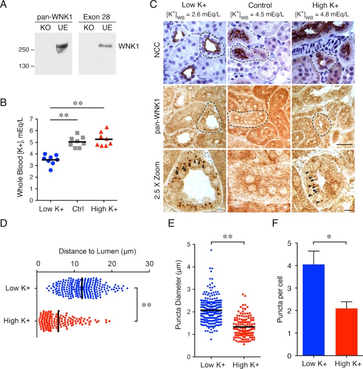
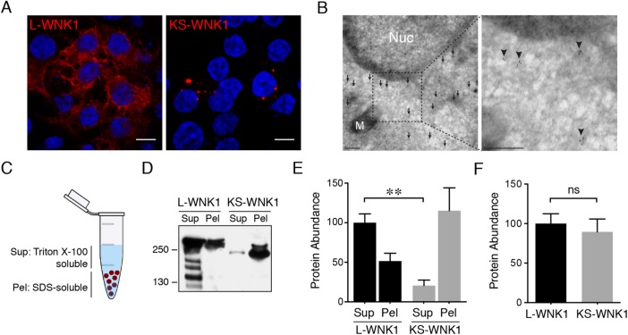
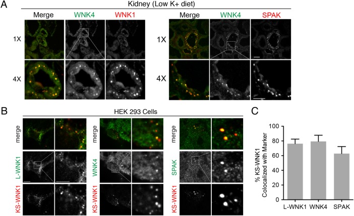
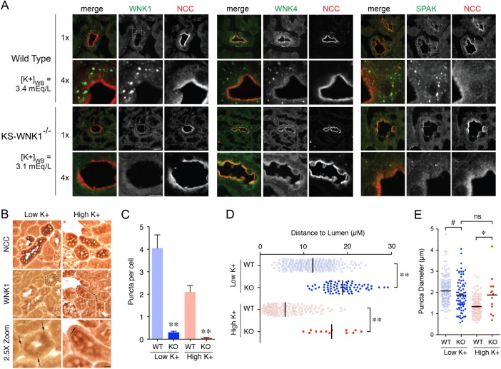
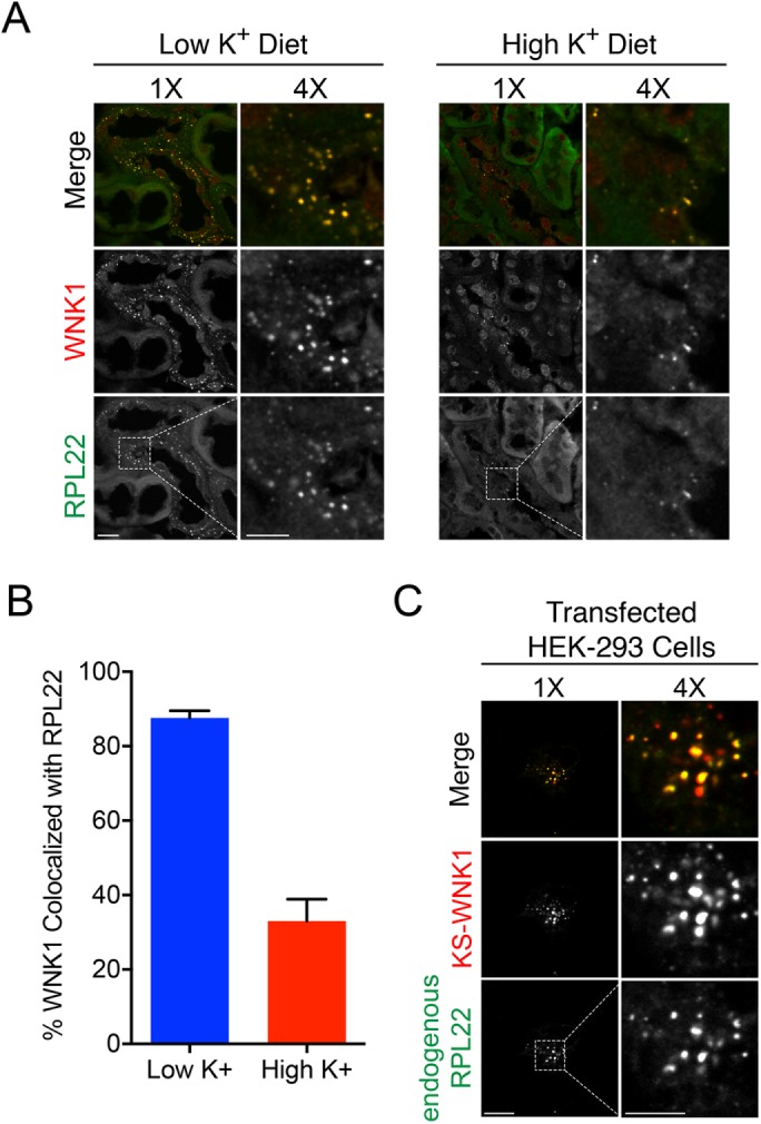
With-no-lysine (WNK) kinases coordinate volume and potassium homeostasis by regulating renal tubular electrolyte transport. In the distal convoluted tubule (DCT), potassium imbalance causes WNK signaling complexes to concentrate into large discrete foci, which we call "WNK bodies." Although these structures have been reported previously, the mechanisms that drive their assembly remain obscure. Here, we show that kidney-specific WNK1 (KS-WNK1), a truncated kinase-defective WNK1 isoform that is highly expressed in the DCT, is critical for WNK body formation. While morphologically distinct WNK bodies were evident in the distal tubules of mice subjected to dietary potassium loading and restriction, KS-WNK1 knockout mice were deficient in these structures under identical conditions. Combining in vivo observations in kidney with reconstitution studies in cell culture, we found that WNK bodies are dynamic membraneless foci that are distinct from conventional organelles, colocalize with the ribosomal protein L22, and cluster the WNK signaling pathway. The formation of WNK bodies requires an evolutionarily conserved cysteine-rich hydrophobic motif harbored within a unique N-terminal exon of KS-WNK1. We propose that WNK bodies are not pathological aggregates, but rather are KS-WNK1-dependent microdomains of the DCT cytosol that modulate WNK signaling during physiological shifts in potassium balance.


Figures

FIGURE 1:
FIGURE 1:
Dietary potassium maneuvers stimulate WNK1 puncta formation in mouse distal convoluted tubules. (A) pan-WNK1 antibody validation using gene-edited WNK1 KO and unedited (UE) cell lines, compared with a previously validated WNK1 antibody against exon 28 (Roy et al., 2015b). (B) Whole blood potassium ([K+]WB) in mice treated with LK, control, or HK diet for 10 d. (n = 8 mice per condition; **: p < 0.0001; ANOVA with Tukey posttest). (C) Representative immunohistochemical staining of kidney tissue from mice on LK, control, or HK diet. [K+]WB, measured by cardiac puncture at the time of kidney harvest, is indicated for each condition. DCTs were identified by NCC/nuclear costaining in contiguous sections. DCT in 2.5× zoom indicated by a dashed line. (n = 5 mice per condition; scale bar = 50 μm in 1× images, 10 μm in 2.5× images). (D–F) Quantification of puncta distance (D), diameter (E), and number per cell (F) under LK and HK conditions (n = 3 mice and more than 59 cells from five tubules per condition; **: p < 0.0001, *: p = 0.02, unpaired t test).
FIGURE 2:
FIGURE 2:
In contrast to L-WNK1, KS-WNK1 forms large puncta in vitro. (A) Immunofluoresence of HEK-293 cells transiently transfected with either L-WNK1-HA or KS-WNK1-HA. (n = 5 transfections; scale bar = 10 μm). (B) Immunogold electron micrographs of HEK-293 cells transiently transfected with KS-WNK1-HA, labeled with anti-HA antibody. Note the concentration of gold particles (arrows) in an electron hypodense region of the cytosol. M = mitochondria; Nuc = nucleus. Scale bar = 100 nm. (C) Supernatant/pellet (SP) assay. Cell lysates were separated into Triton-soluble and Triton-resistant, SDS–soluble fractions. (D) Immunoblots of HEK-293 cells transiently transfected with either L-WNK1-HA or KS-WNK1-HA, subjected to SP assay. Blots were probed with HA antibody revealing a band at ∼250 kDa, corresponding to the MW of L-WNK1 and slightly lower band for KS-WNK1. L-WNK1-HA Sup also contains several other bands, presumably degradation products. (E) Relative protein abunance of L-WNK1 vs. KS-WNK1 in the SP assay. Data were normalized to the L-WNK1 protein abundance in the Sup fraction. (n = 7 transfections; **: p = 0.0021, paired t test). (F) Comparison of the summed supernatant plus pellet protein abunance of L-WNK1 vs. KS-WNK1 in transiently transfected HEK-293 cells (n = 7 transfections; NS by unpaired t test).
FIGURE 3:
FIGURE 3:
WNK1 puncta colocalize with the WNK-SPAK pathway in vivo and in vitro. (A) Immunofluorescence of kidney sections from mice on LK diet × 10 d ([K+]WB = 3.5 mEq/L) costained for WNK4 (both panel sets), pan-WNK1 (left), and SPAK (right). (n = 4 mice per condition; scale bar = 10 μm in 1× images, 5 μm in 4× images). (B) HEK-293 cells were transiently transfected with KS-WNK1-HA and were costained for HA epitopes (all panel sets), transiently transfected myc-L-WNK1 (with anti-myc antibody [left]), endogenous WNK4 (middle), or endogenous SPAK (right) (n = 4 transfections). (C) Percent colocalization in HEK-293 cells of transiently transfected KS-WNK1-HA with exogenous myc-L-WNK (n = 8 images obtained at 60× magnification with an average of four kidney tubules per field), endogenous WNK4 (n = 6 images), or endogenous SPAK (n = 7 images). Pearson correlation coefficients were calculated with Imaris (Bitplane).
FIGURE 4:
FIGURE 4:
The DCT-specific puncta are KS-WNK1 dependent. (A) Immunofluorescence of mouse DCT from either WT or KS-WNK1−/− (KO) mice maintained on LK diet for 10 d. [K+]WB levels were similar for both WT and KS-WNK1 KO mice. DCTs were identified by NCC costaining. Pan-WNK1, WNK4, and SPAK antibodies detected puncta in WT mice, whereas puncta were nearly absent in KS-WNK1 KO mice. (n = 4 mice per genotype; scale bar = 10 μm in 1× images, 5 μm in 4× images). (B) Representative immunohistochemical staining of DCTs from KS-WNK1 KO mice maintained on either LK or HK diet for 10 d. Indicated with arrows are rare punctate structures that were detected in a small subset of DCTs with the pan-WNK1 antibody under both LK and HK conditions. (n = 3 mice per condition; DCT in 2.5× zoom indicated by a dashed line). (C–E) Comparison of WT and KS-WNK1 KO mice on LK and HK diets. KS-WNK1 KO mice exhibited dramatically reduced puncta abundance (C) compared with WT mice. These foci were positioned farther from the lumen (D) and demonstrated a normalization of diameter relative to WT (E; i.e., in KO mice, puncta diameter averaged 1.9 µM under both LK and HK conditions) (n = 3 mice per condition, and due to the scarcity of puncta more than 200 cells from 20 tubules were analyzed per condition. WT data from Figure 1 are presented alongside KO data for comparison. **: p < 0.0001; *: p = 0.0045, #: p = 0.03 by ANOVA, Tukey’s post hoc test).
FIGURE 5:
FIGURE 5:
WNK bodies colocalize with RPL22. (A) Immuno­fluorescence of kidney sections from WT mice on LK or HK diets × 10 d, showing colocalization of pan-WNK1 puncta with RPL22. Box indicates area magnified (bar 1× 10 μm, bar 4× 5 μm). (B) Percent colocalization of between WNK1 and RPL22 under LK and HK dietary conditions. In kidney tissue, on LK diet, WNK1 was 85% colocalized with RPL22, whereas on a HK diet WNK1 was less colocalized with RPL22 (n = 3 mice per condition and 10 images obtained at 60× magnification with an average of four kidney tubules per field). (C) Immunofluorescence of HEK-293 cells transiently transfected with KS-WNK1-HA and costained for HA epitopes and endogenous RPL22 (n = 3 transfections; scale bar = 10 μm in 1× images, 5 μm in 4× images).
FIGURE 6:
FIGURE 6:
A CRH motif localizes KS-WNK1 to WNK bodies. (A) Amino acid sequence of exon 4a. The sequence emerged in coelacanths and is highly conserved to humans, including a putative CRH motif (highlighted in blue). (B) Exon 4a mutagenesis. Five constructs were created including 1) Δ30, lacking the entire exon 4a, 2) C6→S, mutating all of the cysteines to serines, 3) C2→S, mutating the two outer cysteines to serines, 4) C4→S, mutating the four inner cysteines to serines, and 5) 5Φ→5Q, mutating the hydrophobic cluster to glutamines. (C, D) SP assay (anti-HA immunoblot) of HEK-293 cells transiently transfected with KS-WNK1-HA mutants. Representative immunofluorescence of HEK-293 cells transiently transfected with either KS-WNK1-HA or N-terminal mutants, probed with HA antibody (n = 3 transfections). (E) Fluorescence recovery after photobleaching (FRAP) experiment. HEK-293 cells were transiently transfected with L-WNK1-GFP, KS-WNK1-GFP, or C6→S-KS-WNK1-GFP. Representative time series of WNK1-GFP recovery after photobleaching (area bleached circled in blue and recovery indicated by yellow arrows). (F) Percent recovery of the mobile fraction over 20 s. L-WNK1 and the C6→S mutant exhibited a similar rate of recovery (n = 10 replicates for both). In contrast, KS-WNK1 puncta (n = 3 replicates) exhibited limited mobility, with only 5% recovery by 20 s. Error bars = SEM.

References

    1. Al-Qusairi L, Basquin D, Roy A, Stifanelli M, Rajaram RD, Debonneville A, Nita I, Maillard M, Loffing J, Subramanya AR, Staub O. Renal tubular SGK1 deficiency causes impaired K+ excretion via loss of regulation of NEDD4-2/WNK1 and ENaC. Am J Physiol Renal Physiol. 2016:F330–F342. - PMC - PubMed
    1. Al-Qusairi L, Basquin D, Roy A, Rajaram RD, Maillard MP, Subramanya AR, Staub O. Renal tubular ubiquitin-protein ligase NEDD4-2 is required for renal adaptation during long-term potassium depletion. J Am Soc Nephrol. 2017:2431–2442. - PMC - PubMed
    1. Anderson P, Kedersha N. RNA granules. J Cell Biol. 2006:803–808. - PMC - PubMed
    1. Boyd-Shiwarski CR, Subramanya AR. The renal response to potassium stress: integrating past with present. Curr Opin Nephrol Hypertens. 2017:411–418. - PMC - PubMed
    1. Castaneda-Bueno M, Cervantes-Perez LG, Rojas-Vega L, Arroyo-Garza I, Vazquez N, Moreno E, Gamba G. Modulation of NCC activity by low and high K(+) intake: insights into the signaling pathways involved. Am J Physiol Renal Physiol. 2014:F1507–F1519. - PMC - PubMed

Publication types

LinkOut - more resources