. 2017 Dec;20(12):1345-1353.
doi: 10.22038/IJBMS.2017.9622.

The effects of activated omental extract on nuclear and cytoplasmic in vitro maturation of rat oocytes

Affiliations

The effects of activated omental extract on nuclear and cytoplasmic in vitro maturation of rat oocytes

Fakhroddin Mesbah et al. Iran J Basic Med Sci. 2017 Dec.

Abstract

Objective: The role of growth factors, including vascular endothelial growth factor of activated omentum on mitosis is clearly known, though not on all the aspects of in vitro oocyte maturation. This study was designed to assess the effect of activated-omental extract (AOE) on in vitro maturation (IVM) of rat cumulus-oocyte complexes (COCs).

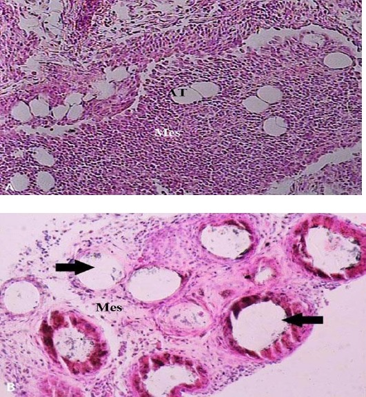
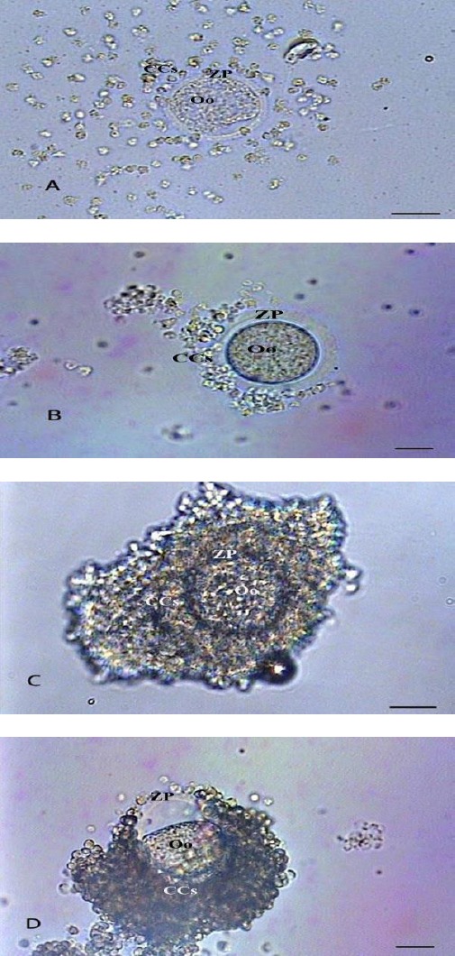
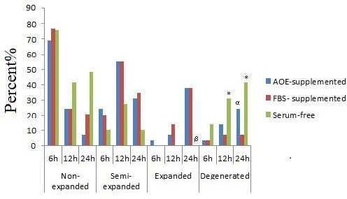
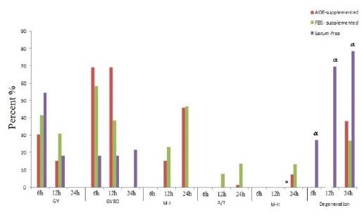
Materials and methods: In this experimental study, the COCs were incubated in Ham's F-10 supplemented with either 20% AOE, 20% fetal bovine serum (FBS) or serum-free media. Post-culture COCs were studied according to the cumulus cells (CCs) expansion, nuclear maturation and cytoplasmic maturation. Cumuli expansion was evaluated by inverted microscope without staining; nuclear maturation was assessed by aceto-orcein staining (light microscope) and cytoplasmic maturation was also observed by TEM.

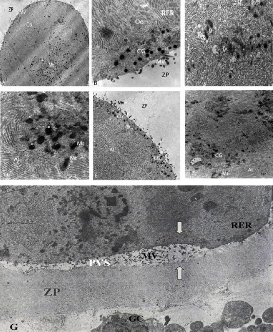
Results: Expansion of CCs and nuclear maturation of the oocytes in in vitro for 24 hr was significantly higher in AOE- and FBS-supplemented groups (P=0.000 and 0.013) and (P=0.004 and 0.014), respectively, compared to serum-free group. At ultra-structural level, after 24 hr, both FBS and AOE-supplemented media showed uniformly wide perivitelline space (PVS). After 12 hr, the cortical granules were found in the oocytes cultured in FBS and AOE-supplemented media. Within 24 hr, both granules and mitochondria were large without any detectable topographic tendency across the ooplasm. In AOE and FBS-supplemented oocytes, the number and size of microvilli were more than those in serum-free one.

Conclusion: Although AOE supplementation induced a higher rate of the CCs expansion, and resuming meiosis, it was not as potent as FBS to provide cytoplasmic maturation of rat oocytes.

Keywords: Cumulus cells; Cytoplasm; In vitro oocyte maturation; Nucleus; Omentum; Rats.


Figures

Figure 1
Figure 1
A) Morphology of a normal omentum. Densely arranged mesothelial cells with sparse adipose cells between them can be observed in a normal omentum micrograph. X150. B) Morphology of an activated omentum. Pathologic cavitation (arrows) indicates the activation of omentum in response to the injection of polydextran particles, which has been washed away during tissue processing. Loosely arranged mesothelial cells with almost no adipose tissue can be observed in an activated omentum micrograph X200. (H & E staining). Mes. (Mesothelium); AT (adipose tissue)
Figure 2
Figure 2
Photomicrographs of post-culture cumulus oocyte complexes at different cumuli expansion levels: expanded (A), semi-expanded (B), non-expanded (C) and degenerated (D) (Bars=5 µm). Oo (oocyte); ZP (zona pellucida); CCs (cumulus cells)
Figure 3
Figure 3
Cumulus cells expansion in AOE- and FBS-supplemented and serum-free groups. AOE; Activated-omental extract, FBS; Fetal bovine serum
Figure 4
Figure 4
Nuclear maturation in AOE- and, FBS-supplemented and serum-free groups. AOE; Activated-omental extract, FBS; Fetal bovine serum, GV: Germinal vesicle; GVBD: Germinal vesicle breakdown: MI: Meiosis I; A/T: Anaphase-telophase: M-II; Meiosis II * Serum-free group versus AOE-supplemented (P=0.014) and FBS- supplemented groups (P=0.004) α Serum-free group versus AOE-supplemented (P=0.014) and FBS- supplemented groups (P<0.001)
Figure 5
Figure 5
Electron micrographs of cytoplasmic characteristics: A) cultured for 12 hr in activated omental extract-supplemented medium (×1200); B) cultured for 24 hr in FBS-supplemented medium (×11300); C) cultured for 6 hr in serum-free medium (×10400); D) cultured for 24 hr in activated omental extract-supplemented medium (stars show CG) (×11000); E) cultured for 24 hr in activated omental extract-supplemented medium (×7500); F) cultured for 6 hr in FBS-supplemented medium (×5800); G) cultured for 24 hr in serum-free medium, arrows show uneven ZP and PVS (×3400). AL (annullate lamellae); CG (cortical granule); GC (granolosa cell); Mt (mitochondria); Mv (microvilli); Oo (oocyte); PVS (perivitelline space); RER (rough endoplasmic reticulum); ZP (zona pellucida)

Similar articles

Cited by

References

    1. Mesbah F, Moslem M, Vojdani Z, Mirkhani H. Does metformin improve in vitro maturation and ultrastructure of oocytes retrieved from estradiol valerate polycystic ovary syndrome-induced rats. J Ovarian Res. 2015;8:874–880. - PMC - PubMed
    1. Von Stetina JR, Orr-Weaver TL. Developmental control of oocyte maturation and egg activation in metazoan models. Cold Spring Harb Perspect Biol. 2011;3:a005553. - PMC - PubMed
    1. Edwards RG. Maturation in vitro of human ovarian oocytes. Lancet. 1965;2:926–929. - PubMed
    1. Ellenbogen A, Shavit T, Shalom-Paz E. IVM results are comparable and may have advantages over standard IVF. Facts View Vis Obgyn. 2014;6:77–80. - PMC - PubMed
    1. Coticchio G, Dal Canto M, Fadini R, Mignini Renzini M, Guglielmo MC, Miglietta S, et al. Ultrastructure of human oocytes after in vitro maturation. Mol Hum Reprod. 2016;22:110–118. - PubMed

LinkOut - more resources