Review
. 2019 Jan;286(2):379-398.
doi: 10.1111/febs.14358. Epub 2017 Dec 29.

ER stress in skeletal muscle remodeling and myopathies

Affiliations
Review

ER stress in skeletal muscle remodeling and myopathies

Dil Afroze et al. FEBS J. 2019 Jan.

Abstract

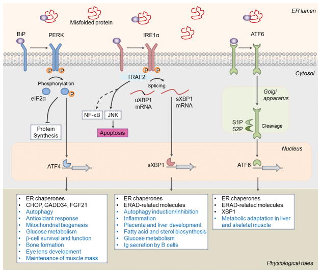
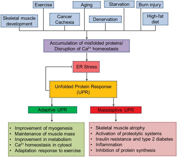
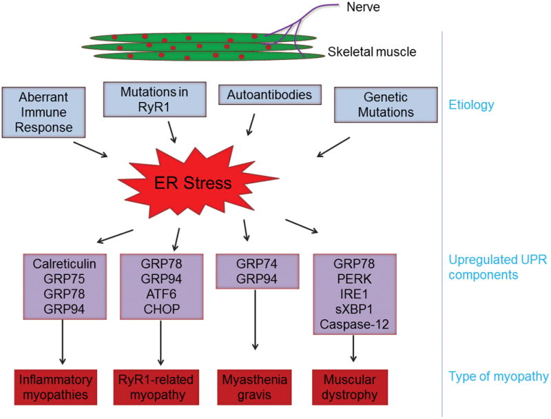
Skeletal muscle is a highly plastic tissue in the human body that undergoes extensive adaptation in response to environmental cues, such as physical activity, metabolic perturbation, and disease conditions. The endoplasmic reticulum (ER) plays a pivotal role in protein folding and calcium homeostasis in many mammalian cell types, including skeletal muscle. However, overload of misfolded or unfolded proteins in the ER lumen cause stress, which results in the activation of a signaling network called the unfolded protein response (UPR). The UPR is initiated by three ER transmembrane sensors: protein kinase R-like endoplasmic reticulum kinase, inositol-requiring protein 1α, and activating transcription factor 6. The UPR restores ER homeostasis through modulating the rate of protein synthesis and augmenting the gene expression of many ER chaperones and regulatory proteins. However, chronic heightened ER stress can also lead to many pathological consequences including cell death. Accumulating evidence suggests that ER stress-induced UPR pathways play pivotal roles in the regulation of skeletal muscle mass and metabolic function in multiple conditions. They have also been found to be activated in skeletal muscle under catabolic states, degenerative muscle disorders, and various types of myopathies. In this article, we have discussed the recent advancements toward understanding the role and mechanisms through which ER stress and individual arms of the UPR regulate skeletal muscle physiology and pathology.

Keywords: ER stress; metabolism; myogenesis; myopathies; skeletal muscle atrophy; unfolded protein response.


Conflict of interest statement

Conflict of interest statement: The authors report no conflicts of interest.

Figures

FIGURE 1
FIGURE 1. Activation and mode of action of the unfolded protein response (UPR) in physiological processes
In absence of stress, endoplasmic reticulum (ER) chaperone protein, BiP/GRP78, binds to the UPR sensors: protein kinase R (PKR)-like endoplasmic reticulum kinase (PERK), inositol-requiring protein 1α (IRE1α), and activating transcription factor 6 (ATF6) to keep them inactive. Upon ER stress, BiP disassociates from these sensors and preferentially binds to the misfolded proteins in the ER lumen. Upon release from BiP, PERK undergoes dimerization and autophosphorylation leading to a cascade of signals including phosphorylation of eIF2α, inhibition of protein synthesis, and selective upregulation of translation of ATF4, a potent transcription factor. The PERK arm of the UPR also regulates mitochondrial biogenesis, antioxidant response, and gene expression of C/EBP homologous protein (CHOP), growth arrest and DNA damage-inducible protein 34 (GADD34), fibroblast growth factor 21 (FGF21), and enzymes involved in amino acid metabolism. IRE1α also becomes activated by dimerization and autophosphorylation during ER stress. Through its endonuclease activity, IRE1α processes the mRNA encoding unspliced XBP1 (uXBP1) to produce an active transcription factor, spliced XBP1 (sXBP1). The sXBP1 controls the transcription of genes encoding ER chaperones, ER-associated protein degradation (ERAD), and protein quality control and phospholipid synthesis. Through interacting with adaptor proteins, such as TNF receptor-associated protein 2 (TRAF2), IRE1α activates c-Jun N-terminal kinase (JNK) and potentially nuclear factor-kappa B (NF-κB) pathways to regulate cell fate. Upon ER stress, ATF6 is transported to the Golgi apparatus, where it is processed by site 1 protease (S1P) and S2P, releasing its cytosolic domain fragment which translocate to the nucleus to induce gene expression of components of ERAD as well as XBP1. Studies using genetic mouse models have revealed that individual arm of the UPR may regulate common or distinct physiological response as depicted here.
FIGURE 2
FIGURE 2. Involvement of ER stress and the UPR in skeletal muscle remodeling
Many physiological stimuli and disease states lead to the activation of ER stress-induced UPR pathways. Although genetic evidence is still sparse, the UPR can improve or deteriorate skeletal muscle mass and function. Adaptive UPR generally improves skeletal muscle formation and metabolic function and promotes maintenance of skeletal muscle mass during exercise and other perturbation. Adaptive UPR also inhibits ER stress through improving protein folding and restoring Ca2+ homeostasis in ER. Chronic activation of UPR can lead to skeletal muscle wasting through repressing the rate of protein synthesis, activation of proteolytic pathways, inflammation, and development of insulin resistance.
FIGURE 3
FIGURE 3. Role of ER stress in myopathies
Chronic activation of ER stress occurs in several types of myopathies. The underlying cause and various components of ER stress and the UPR found to be activated in different types of myopathy are summarized here. RyR1, Ryanodine receptor type 1.

References

    1. Pedersen BK, Febbraio MA. Muscles, exercise and obesity: skeletal muscle as a secretory organ. Nat Rev Endocrinol. 2012;8:457–65. - PubMed
    1. Egerman MA, Glass DJ. Signaling pathways controlling skeletal muscle mass. Crit Rev Biochem Mol Biol. 2014;49:59–68. - PMC - PubMed
    1. Glass DJ. Skeletal muscle hypertrophy and atrophy signaling pathways. Int J Biochem Cell Biol. 2005;37:1974–84. - PubMed
    1. Shin JY, Mendez-Lopez I, Wang Y, Hays AP, Tanji K, Lefkowitch JH, Schulze PC, Worman HJ, Dauer WT. Lamina-associated polypeptide-1 interacts with the muscular dystrophy protein emerin and is essential for skeletal muscle maintenance. Dev Cell. 2013;26:591–603. - PMC - PubMed
    1. Hetz C, Chevet E, Oakes SA. Proteostasis control by the unfolded protein response. Nat Cell Biol. 2015;17:829–38. - PMC - PubMed

Publication types