. 2018 Jan;7(1):208-218.
doi: 10.1002/cam4.1269. Epub 2017 Dec 13.

Oxymatrine inhibits non-small cell lung cancer via suppression of EGFR signaling pathway

Affiliations

Oxymatrine inhibits non-small cell lung cancer via suppression of EGFR signaling pathway

Wei Li et al. Cancer Med. 2018 Jan.

Abstract

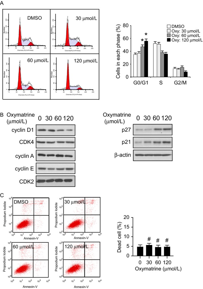
Epidermal growth factor receptor (EGFR) plays a crucial role in human non-small cell lung cancer (NSCLC) tumorigenesis. In this study, oxymatrine was identified as an EGFR signaling pathway inhibitor in NSCLC. Oxymatrine inhibited anchorage-dependent and independent growth of NSCLC cell lines but had no cytotoxicity in normal lung cells. We found that exposure to oxymatrine not only suppressed the activity of wild-type EGFR but also inhibited the activation of exon 19 deletion and L858R/T790M mutated EGFR. Flow cytometry analysis suggested that oxymatrine-induced cell cycle G0/G1 arrest was dependent on EGFR-Akt signaling. Exogenous overexpression of Myr-Akt rescued cyclin D1 expression in HCC827 cells. Moreover, oxymatrine prominently suppressed tumor growth in a xenograft mouse model. Thus, oxymatrine appears to be a novel therapeutic agent for NSCLC treatment.

Keywords: Akt; cyclin D1; epidermal growth factor receptor; non-small cell lung cancer; oxymatrine.


Figures

Figure 1
Figure 1
The structure and cytotoxicity of oxymatrine. (A) The chemical structure of oxymatrine. (B) Cytotoxicity of oxymatrine was measured in normal lung cells by MTS assay. MRC‐5, NL20, and HBE cells were treated with various concentrations of oxymatrine for 24 h. Data are shown as means ± SD from triplicate experiments.
Figure 2
Figure 2
Inhibitory effects of oxymatrine on NSCLC cells. (A) Oxymatrine inhibits anchorage‐dependent cell growth in a panel of human NSCLC cells, including A549 (left), H1975 (middle), and HCC827 (right). Cell viability was measured by MTS assay. (B) Three NSCLC cell lines, including A549 (upper), H1975 (middle), and HCC827 (bottom) were subjected to the soft agar assay. The average colony number was calculated from three separate experiments. Asterisk, significant (*< 0.05, **< 0.01, ***< 0.001) suppression of cell viability or colony formation by oxymatrine compared with the DMSO‐treated group.
Figure 3
Figure 3
Oxymatrine suppresses EGFR signaling pathway. (A) Oxymatrine inhibits the activity of EGFR signaling pathway. A549 (left), H1975 (middle), and HCC827 (right) cells were treated with oxymatrine for 24 h as indicated, Western blot was conducted to detect target proteins. (B) Oxymatrine downregulates EGF‐induced EGFR signaling pathway activation. HCC827 cells were starved overnight and then treated with oxymatrine at the indicated concentrations for 2 h. After stimulation with EGF (0, 50 ng/mL) for 30 min, the cells were harvested and protein levels were determined by Western blotting. (C) Oxymatrine downregulates EGF‐induced EGFR signaling pathway activation. HCC827 cells were starved overnight and then treated with 60 μmol/L oxymatrine for 2 h. After stimulation with EGF (50 ng/mL) for various time points, the cells were harvested and protein levels were determined by Western blotting.
Figure 4
Figure 4
Oxymatrine induces G0/G1 cell cycle arrest in HCC827 cells. (A) HCC827 cells were treated with various concentrations of oxymatrine for 24 h as indicated, flow cytometry was conducted to analyze cell cycle distribution (*P < 0.05 vs. DMSO‐treated). (B) HCC827 cells were treated with various concentrations of oxymatrine for 24 h as indicated, Western blot was conducted to detect target proteins. (C) HCC827 cells were treated with various concentrations of oxymatrine for 24 h as indicated, the percentage of apoptosis cells was quantified by flow cytometry (#, not statistically significant).
Figure 5
Figure 5
Akt inhibition is required for oxymatrine‐mediated cell cycle arrest. (A) HCC827 cells were treated with oxymatrine or inhibitors for 24 h as indicated, protein levels were determined by Western blotting. (B) Overexpression of constitutively activated Akt (Myr‐Akt1) rescues cyclin D1 expression. The Myr‐Akt1 plasmid was transfected into HCC827 cells, after 24 h, these cells were treated with oxymatrine for another 24 h as indicated. Western blot analysis was performed to detect the protein expression levels. (C) Overexpression of Myr‐Akt1 rescues cell cycle arrest in oxymatrine‐treated HCC827 cells. The Myr‐Akt1 plasmid transfection and oxymatrine treatment were performed as indicated. Cell cycle distribution was analyzed by flow cytometry (*P < 0.05 vs. oxymatrine‐treated).
Figure 6
Figure 6
Oxymatrine inhibits tumor growth in HCC827 xenograft mouse model. (A) HCC827 cells were subcutaneously injected into the right flank of mice. At the treatment endpoint, mice were killed and tumors were removed, weighed, and photographed. (B) Tumor volumes were measured twice a week. (C) The tumor weight from the vehicle‐ and oxymatrine‐treated group was measured. (D) During the treatment period, the body weight of mice was measured twice a week to determine the effect of oxymatrine. For (B), (C), and (D), data are shown as mean values ± SD obtained from five mice in each group. (E) Immunohistochemical staining examination of Ki67 and p‐EGFR in tumor sections from the vehicle‐ or the oxymatrine‐treated group. The integrated optical density (IOD) was evaluated using the Image‐Pro Plus software (version 6.2) program. (*< 0.05, **< 0.01 vs. vehicle).

Similar articles

Cited by

References

    1. Siegel, R. , Naishadham D., and Jemal A.. 2013. Cancer statistics, 2013. CA Cancer J. Clin. 63:11–30. - PubMed
    1. DeSantis, C. E. , Lin C. C., Mariotto A. B., Siegel R. L., Stein K. D., Kramer J. L., et al. 2014. Cancer treatment and survivorship statistics, 2014. CA Cancer J. Clin. 64:252–271. - PubMed
    1. Thomas, A. , Liu S. V., Subramaniam D. S., and Giaccone G.. 2015. Refining the treatment of NSCLC according to histological and molecular subtypes. Nat. Rev. Clin. Oncol. 12:511–526. - PubMed
    1. Rosell, R. , and Karachaliou N.. 2015. Lung cancer in 2014: optimizing lung cancer treatment approaches. Nat. Rev. Clin. Oncol. 12:75–76. - PubMed
    1. Herbst, R. S. , Heymach J. V., and Lippman S. M.. 2008. Lung cancer. N. Engl. J. Med. 359:1367–1380. - PMC - PubMed

MeSH terms