

Magnetic resonance elastography for examining developmental changes in the mechanical properties of the brain
- PMID: 29239832
- PMCID: PMC5832528
- DOI: 10.1016/j.dcn.2017.08.010
Magnetic resonance elastography for examining developmental changes in the mechanical properties of the brain
Abstract
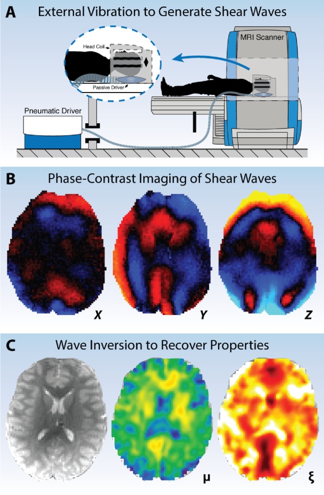
Magnetic resonance elastography (MRE) is a quantitative imaging technique for noninvasively characterizing tissue mechanical properties, and has recently emerged as a valuable tool for neuroimaging. The measured mechanical properties reflect the microstructural composition and organization of neural tissue, and have shown significant effects in many neurological conditions and normal, healthy aging, and evidence has emerged supporting novel relationships between mechanical structure and cognitive function. The sensitivity of MRE to brain structure, function, and health make it an ideal technique for studying the developing brain; however, brain MRE studies on children and adolescents have only just begun. In this article, we review brain MRE and its findings, discuss its potential role in developmental neuroimaging, and provide suggestions for researchers interested in adopting this technique.
Keywords: Brain; Development; Elastography; Stiffness; Viscoelasticity.
Copyright © 2017 The Authors. Published by Elsevier Ltd.. All rights reserved.
Figures
References
-
- Braun J., Guo J., Lutzkendorf R., Papazoglou S., Hirsch S., Sack I., Bernarding J. High-resolution mechanical imaging of the human brain by three-dimensional multifrequency magnetic resonance elastography at 7T. Neuroimage. 2014;90:308–314. - PubMed
-
- Deary I.J., Bastin M.E., Pattie A., Clayden J.D., Whalley L.J., Starr J.M., Wardlaw J.M. White matter integrity and cognition in childhood and old age. Neurology. 2006;66:505–512. - PubMed
Publication types
MeSH terms
Grants and funding
LinkOut - more resources
Full Text Sources
Other Literature Sources
