. 2018 Mar 16;13(3):582-590.
doi: 10.1021/acschembio.7b00685. Epub 2018 Jan 3.

Structural Basis of ALDH1A2 Inhibition by Irreversible and Reversible Small Molecule Inhibitors

Affiliations

Structural Basis of ALDH1A2 Inhibition by Irreversible and Reversible Small Molecule Inhibitors

Yan Chen et al. ACS Chem Biol. .

Abstract

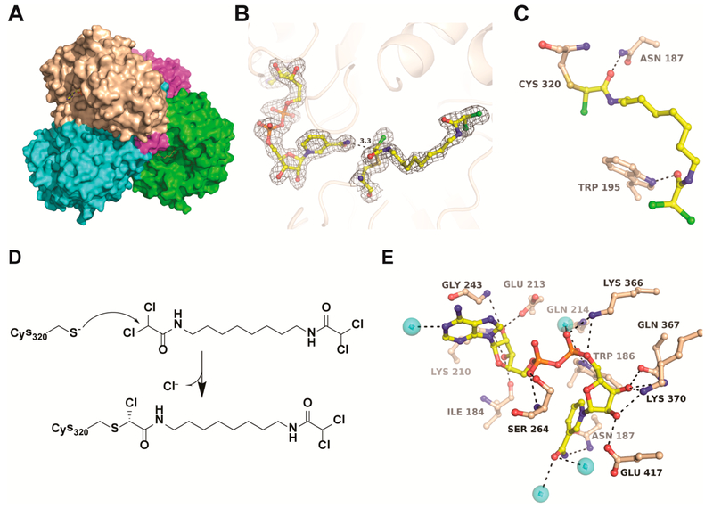
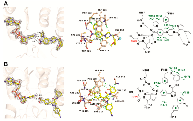
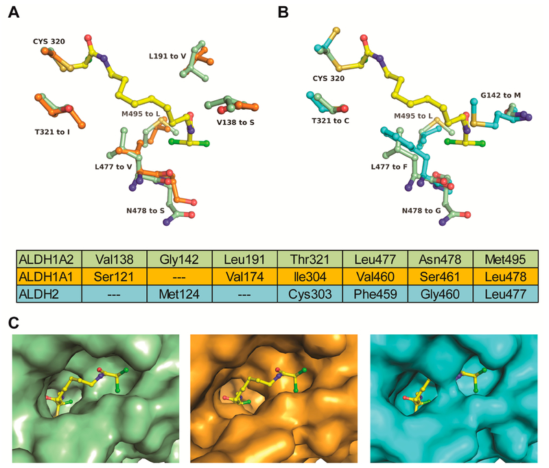
Enzymes of the ALDH1A subfamily of aldehyde dehydrogenases are crucial in regulating retinoic acid (RA) signaling and have received attention as potential drug targets. ALDH1A2 is the primary RA-synthesizing enzyme in mammalian spermatogenesis and is therefore considered a viable drug target for male contraceptive development. However, only a small number of ALDH1A2 inhibitors have been reported, and information on the structure of ALDH1A2 was limited to the NAD-liganded enzyme void of substrate or inhibitors. Herein, we describe the mechanism of action of structurally unrelated reversible and irreversible inhibitors of human ALDH1A2 using direct binding studies and X-ray crystallography. All inhibitors bind to the active sites of tetrameric ALDH1A2. Compound WIN18,446 covalently reacts with the side chain of the catalytic residue Cys320, resulting in a chiral adduct in ( R) configuration. The covalent adduct directly affects the neighboring NAD molecule, which assumes a contracted conformation suboptimal for the dehydrogenase reaction. The reversible inhibitors interact predominantly through direct hydrogen bonding interactions with residues in the vicinity of Cys320 without affecting NAD. Upon interaction with inhibitors, a large flexible loop assumes regular structure, thereby shielding the active site from solvent. The precise knowledge of the binding modes provides a new framework for the rational design of novel inhibitors of ALDH1A2 with improved potency and selectivity profiles.


Figures

Figure 1.
Figure 1.
Binding potential and activity of ALDH1A2 inhibitors. (A) Chemical structures of the inhibitors studied in this work. (B) Direct binding studies by ITC. The top panels show the raw titration data; the bottom panels show the binding isotherms. (C) Thermodynamic signature of ALDH1A2 interaction with NAD and inhibitors. (D) Dose-dependent inhibition of the ALDH1A2 reaction with retinal and NAD as substrate and cofactor. The dotted lines show the activity of WIN18,446 upon different preincubation times with the enzyme prior to starting the reaction with retinal. The Kd and IC50 values are listed in Table 1.
Figure 2.
Figure 2.
Crystal structure of the ALDH1A2-WIN18,446 complex. (A) Surface presentation of the ALDH1A2–WIN18,446 tetramer with each polypeptide chain colored differently. (B) 2FoFc electron density around NAD and WIN18,446 contoured at 1σ. Electron density maps of inhibitor in all four polypeptide chains are shown in Supporting Information Figure S2. (C) The covalent adduct between Cys320 and WIN18,446 along with H-bonding interactions in the active site. A stereo presentation of all binding interactions is shown in Supporting Information Figure S3. (D) Proposed mechanism for the covalent reaction of WIN18,446 with Cys320. (E) Hydrogen bonding interactions of NAD with residues of the cofactor site. Black dotted lines indicate H-bonding interactions.
Figure 3.
Figure 3.
Crystal structures of ALDH1A2 in complex with reversible inhibitors. (A) ALDH1A2 in complex with NAD and compound 6–118. (B) ALDH1A2 in complex with NAD and compound CM121. The left panels show the 2FoFc electron density map contoured at 1σ around NAD and inhibitor. The middle panels show potential H-bonding (black dotted lines) and VDW interactions (green dotted lines) of the inhibitors in the active site. The right panels show a schematic drawing of the inhibitor interactions. Electron density maps of inhibitor in all four polypeptide chains are shown in Supporting Information Figure S2. Stereo presentations of the binding interactions are shown in Supporting Information Figure S3.
Figure 4.
Figure 4.
Structural consequence of ALDH1A2 interaction with reversible and irreversible inhibitors. (A) Root mean square deviation (RMSD) of the Cα atoms of ALDH1A2 (chain A) liganded with compound 6–118 (blue) and CM121 (red) superimposed onto the ALDH1A2–WIN18,446 complex. (B) Superimposed inhibitors WIN18,446 (green), 6–118 (yellow), and CM121 (magenta) upon alignment of the respective ALDH1A2 cocrystal structures. The inhibitor binding site extends ~9 Å from Cys320 in the interior of the protein toward the solvent exposed area. A hydrophobic subpocket accommodates halogen-containing moieties of WIN18,446 and CM121. (C) Formation of the covalent adduct of Cys320 with WIN18,446 results in a steric clash with the neighboring nicotinamide group of NAD in its extended conformation (yellow). As a consequence, NAD assumes a contracted conformation (green). (D) The contracted conformation of NAD in the ALDH1A2–WIN18,446 complex (green) is similar to that of NADH in ALDH2 (PDB entry 1NZW, cyan). (E) Extended conformation of NAD as observed in the structures of ALDH1A2 with 6–118 (yellow) and CM121 (magenta).
Figure 5.
Figure 5.
A large flexible loop in ALDH1A2 assumes regular structure upon inhibitor binding. (A) Monomeric human ALDH1A2 with partially occupied NAD (PDB 4X2Q, cyan) superimposed on the ALDH1A2–Win18,446 complex (beige). A 21 residue loop flanking the substrate binding site is flexible in the uninhibited enzyme but rigidifies in the dead-end complex (magenta). (B) 2FoFc electron density of the loop residues contoured at 1σ; the corresponding amino acid sequence is also shown. (C) Intermolecular contacts of tetrameric ALDH1A2 between the 475–495 loop of one monomer (magenta) with the 162–166 loop of a neighboring monomer (green). WIN18,446 is shown in yellow.
Figure 6.
Figure 6.
Comparison of the inhibitor binding sites of aldehyde dehydrogenases. (A) Superimposition of the ALDH1A2-WIN18,446 complex (green) with ALDH1A1 (PDB entry 4WPN, orange) shows six residues of the inhibitor binding site that differ between the two enzymes WIN18,446 is shown in yellow. (B) Superimposition with ALDH2 (PDB entry 2VLE, cyan) shows five amino acid substitutions, Gly142Met and Leu477Phe narrowing the binding site considerably. Aligned residues comprising the respective active sites are tabulated. (C) Surface presentations illustrating the effect of amino acid substitutions on the active site architecture of the respective enzymes. WIN18,446 was overlaid onto the ALDH1A1 and ALDH2 structures.

References

    1. Koppaka V, Thompson DC, Chen Y, Ellermann M, Nicolaou KC, Juvonen RO, Petersen D, Deitrich RA, Hurley TD, and Vasiliou V (2012) Aldehyde dehydrogenase inhibitors: a comprehensive review of the pharmacology, mechanism of action, substrate specificity, and clinical application. Pharmacol. Rev 64, 520–539. - PMC - PubMed
    1. Clark DW, and Palle K (2016) Aldehyde dehydrogenases in cancer stem cells: potential as therapeutic targets. Ann. Transl Med. 4, 518. - PMC - PubMed
    1. Liu X, Wang L, Cui W, Yuan X, Lin L, Cao Q, Wang N, Li Y, Guo W, Zhang X, Wu C, and Yang J (2016) Targeting ALDH1A1 by disulfiram/copper complex inhibits non-small cell lung cancer recurrence driven by ALDH-positive cancer stem cells. Oncotarget 7, 58516–58530. - PMC - PubMed
    1. Perez-Alea M, McGrail K, Sanchez-Redondo S, Ferrer B, Fournet G, Cortes J, Munoz E, Hernandez-Losa J, Tenbaum S, Martin G, Costello R, Ceylan I, Garcia-Patos V, and Recio JA (2017) ALDH1A3 is epigenetically regulated during melanocyte transformation and is a target for melanoma treatment. Oncogene 36, 5695–5708. - PubMed
    1. Tomita H, Tanaka K, Tanaka T, and Hara A (2016) Aldehyde dehydrogenase 1A1 in stem cells and cancer. Oncotarget 7, 11018–11032. - PMC - PubMed

Publication types