. 2018 Jan 23;69(3):643-654.
doi: 10.1093/jxb/erx424.

MAP kinases associate with high molecular weight multiprotein complexes

Affiliations

MAP kinases associate with high molecular weight multiprotein complexes

Carlton J Bequette et al. J Exp Bot. .

Abstract

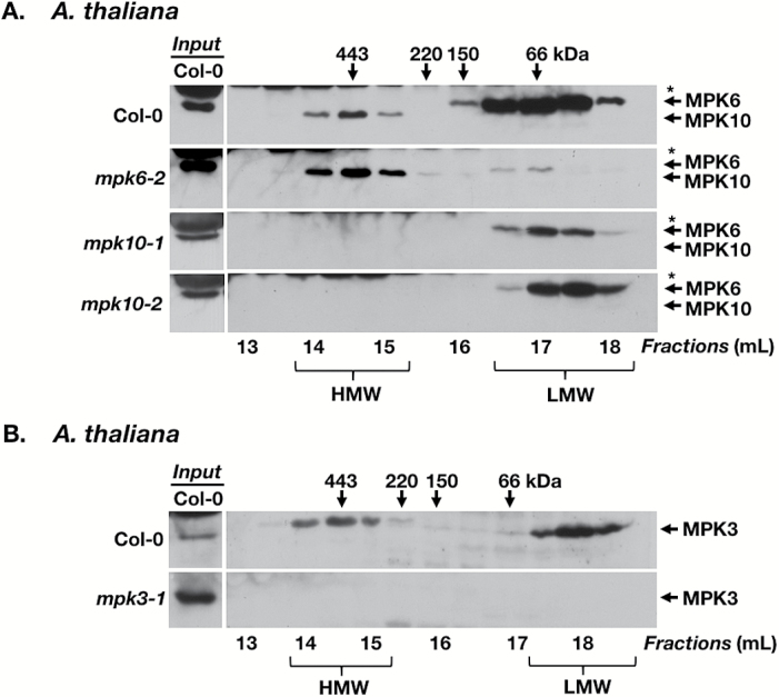
Plant responses to the environment and developmental processes are mediated by a complex signaling network. The Arabidopsis thaliana mitogen-activated protein kinases (MAPKs) MPK3 and MPK6 and their orthologs in other plants are shared signal transducers that respond to many developmental and environmental signals and thus represent highly connected hubs in the cellular signaling network. In animals, specific MAPK signaling complexes are assembled which enable input-specific protein-protein interactions and thus specific signaling outcomes. In plants, not much is known about such signaling complexes. Here, we report that MPK3, MPK6, and MPK10 orthologs in tomato, tobacco, and Arabidopsis as well as tomato MAPK kinase 4 (MKK4) associate with high molecular weight (~250-550 kDa) multiprotein complexes. Elicitation by the defense-associated peptides flg22 and systemin resulted in phosphorylation and activation of the monomeric MAPKs, whereas the complex-associated MAPKs remained unphosphorylated and inactive. In contrast, treatment of tomato cells with a phosphatase inhibitor resulted in association of phosphorylated MPK1/2 with the complex. These results demonstrate that plant MAPKs and MAPKKs dynamically assemble into stable multiprotein complexes and this may depend on their phosphorylation status. Identification of the constituents of these multiprotein complexes promises a deeper understanding of signaling dynamics.

Keywords: Arabidopsis; MAP kinase signaling; MAPK; MAPKK; multiprotein complex; phosphorylation; scaffold; signal transduction; size-exclusion chromatography; tomato.


Figures

Fig. 1.
Fig. 1.
Identification of a MAP kinase-containing multiprotein complex in tomato and tobacco. Total protein was extracted from leaf tissue and separated by GF. Eluate was collected in 0.5 ml fractions, concentrated, and analyzed by immunoblotting (IB). Only fractions representing 13–18.5 ml are shown (numbers underneath the immunoblots). The numbers above the panels indicate the peak elution of molecular weight standards in kDa. Input samples represent 30 μg of total protein extracted from leaf tissue and represent the same protein extracts that were used for GF. (A) GF/IB analysis of leaf extracts (1.5 mg of total protein) from tomato plants (S. lycopersicum). SlMPK1/2 were detected using anti-AtMPK6 antibody. (B) GF/IB analysis of leaf extracts (1.5 mg of total protein) from tomato plants in which MPK1, MPK2, and MPK3 were co-silenced via VIGS (VIGS-mpk1/2/3) and from control plants. SlMPK1/2 were detected using anti-AtMPK6 antibody. Inputs represent 30 µg of total protein from control plants (C) or VIGS-mpk1/2/3 plants (VIGS). (C) GF/IB analysis of leaf extracts (1.2 mg of total protein) from tobacco plants (N. tabacum). SIPK and Ntf4 were detected using anti-AtMPK6 antibody. (D) GF/IB analysis of leaf extracts (1 mg of total protein) from N. benthamiana plants transiently overexpressing 35S:SlMPK1-c-myc or 35S:SlMPK2-c-myc, probed with anti-c-myc antibody. Inputs represent 30 μg of total protein from non-infiltrated N. benthamiana leaves (WT) or from leaves overexpressing fusion proteins (MPK-c-myc). The shift towards higher molecular weight (up to 150 kDa) in the LMW fractions is probably an artifact caused by the high amounts of tagged MPK1/2 protein due to overexpression. HMW, MAPK-containing high molecular weight fractions; LMW, MAPK-containing low molecular weight fractions. (A) is representative of seven, (B) of three, (C) of four, and (D) of one independent experiment, with each sample containing tissue from at least two different plants. *Unrelated protein that cross-reacts with anti-AtMPK6 antibodies.
Fig. 2.
Fig. 2.
Identification of a MAP kinase-containing multiprotein complex in Arabidopsis thaliana. (A) GF/IB analysis of leaf extracts from A. thaliana ecotype Columbia-0 (Col-0, 1.5 mg of total protein), and the null mutants mpk6-2 (1.5 mg), mpk10-1 (0.5 mg), and mpk10-2 (0.5 mg). AtMPK6 and AtMPK10 were detected using anti-AtMPK6 antibody. (B) GF/IB analysis of leaf extracts from A. thaliana ecotype Col-0 (1.5 mg of total protein) and the mpk3-1 null mutant (0.75 mg). AtMPK3 was detected using anti-AtMPK3 antibody. Note: the Col-0 column on the left shows extracts (30 µg of total protein) from Col-0 as a reference for MPK6 (A) or MPK3 (B). These samples were analyzed on the same blot as the corresponding GF fractions, but additional bands were removed to adjust the elution profiles among panels. This is indicated by the white line that separates reference samples and GF fractions. For the two mpk-10 rows, shorter exposures for the Col-0 reference lane are shown as compared with the GF fractions. Numbers above and below the immunoblots are as described in Fig. 1. Immunoblots for Col-0 and mpk6-2 represent four independent experiments; and those for the two independent mpk10 mutant lines were performed once for each line.
Fig. 3.
Fig. 3.
Solanum lycopersicum MKK4 associates with a multiprotein complex. (A) Extracts from N. tabacum (Nt) and S. lycopersicum (Sl) leaves as well as affinity-purified recombinant SlMKK4 protein (RP) were analyzed by IB using either anti-SlMKK4 antiserum (left panel) or anti-SlMKK4 antiserum pre-incubated with recombinant MKK4 protein (right panel). Note that the lane showing purified MKK4 protein contains MKK4 degradation products. The binding of free MKK4 by the antibody prevented it from recognizing MKK4 on the immunoblot membrane. The MKK4 ortholog from N. tabacum (MKK9; accession no. NP_001311802) shares 88% identity with SlMKK4 at the amino acid level. Without a stretch of 16 amino acids that is absent in NtMKK9, the identity is 93%. This explains why the anti-SlMKK4 antibody recognizes a protein in N. tabacum, presumably NtMKK9. (B) GF/IB analysis of leaf extracts (1.5 mg) from S. lycopersicum plants. SlMKK4 was detected using anti-SlMKK4 antibody. Numbers above and below the immunoblots are as described in Fig. 1. All immunoblots represent a minimum of two independent experiments.
Fig. 4.
Fig. 4.
Active phosphorylated MPK1/SIPK and MPK2/Ntf4 do not normally associate with a multiprotein complex in tomato and tobacco. Solanum peruvianum suspension-cultured cells were treated with 10 nM flg22 or 10 nM systemin. Total protein was extracted from suspension cells 0 and 10 min after elicitor treatment. Inputs represent 30 μg of total protein extracted from cells collected at the times indicated after treatment. Numbers above and below immunoblots are as described in Fig. 1. (A) GF/IB analysis of extracts (0.9 mg of total protein) from untreated suspension cells or cells treated with flg22, probed with anti-AtMPK6 antibody for the detection of total MPK1/2 and anti-pERK antibody for the detection of phosphorylated MPK1/2 (p-MPK1/2). (B) The same as (A) but after treatment with systemin (0.75 mg of total protein). The additional panel shows enzymatic activity of MPK1/2 as determined by in-gel kinase assays (IGKAs). Total protein (1.5 mg) was separated by GF and each fraction was analyzed by IGKA. Signals represent 32P-phosphorylated myelin basic protein. (C) GF/IB analysis of extracts (1.2 mg of total protein) from tobacco leaves left untreated or wounded and sampled 10 min later, probed with anti-AtMPK6 antibody for the detection of SIPK/Ntf4 and anti-pERK antibody for the detection of phosphorylated SIPK/Ntf4 (p-SIPK/Ntf4). (A) and immunoblots in (B) are representative of four independent experiments; IGKAs in (B) and (C) of two independent experiments. For tobacco, similar results were seen for 180 min after wounding (Supplementary Fig. S5B). *Unrelated protein that cross-reacts with anti-AtMPK6 antibodies.
Fig. 5.
Fig. 5.
Phosphorylated MPK1 and MPK2 associate with a multiprotein complex after treatment with a phosphatase inhibitor. Solanum peruvianum cells were treated with the phosphatase inhibitor cantharidin and sampled at the times indicated. (A) IB analysis of cell extracts (30 µg) from suspension cells sampled at the times indicated after 500 μM cantharidin treatment, probed with anti-pERK antibody (upper panel) and anti-AtMPK6 antibody (lower panel) for the detection of phosphorylated and total MPK1/2, respectively. (B) GF/IB analysis of cell extracts (1.5 mg total protein) from suspension cells sampled at 360 min after 500 μM cantharidin treatment, probed with anti-pERK antibody for the detection of phosphorylated MPK1/2. Input represents 30 μg of total protein. (C) GF/IB analysis of cell extracts (1.5 mg of total protein) from suspension cells sampled at 10, 120, and 180 min after treatment with 100 μM cantharidin, probed with anti-pERK antibody for the detection of phosphorylated MPK1/2 (upper panel). Immunoblots were stripped and reprobed with anti-AtMPK6 antibody for the detection of total MPK1/2 (lower panel). Only HMW fractions were analyzed (13–15 ml). Numbers above and below the immunoblots are as described in Fig. 1. Similar results were obtained in at least four independent experiments.

References

    1. Alonso JM, Stepanova AN, Leisse TJ, et al. . 2003. Genome-wide insertional mutagenesis of Arabidopsis thaliana. Science 301, 653–657. - PubMed
    1. Andreasson E, Ellis B. 2010. Convergence and specificity in the Arabidopsis MAPK nexus. Trends in Plant Science 15, 106–113. - PubMed
    1. Bartels S, Anderson JC, González Besteiro MA, Carreri A, Hirt H, Buchala A, Métraux JP, Peck SC, Ulm R. 2009. MAP kinase phosphatase1 and protein tyrosine phosphatase1 are repressors of salicylic acid synthesis and SNC1-mediated responses in Arabidopsis. The Plant Cell 21, 2884–2897. - PMC - PubMed
    1. Bartels S, González Besteiro MA, Lang D, Ulm R. 2010. Emerging functions for plant MAP kinase phosphatases. Trends in Plant Science 15, 322–329. - PubMed
    1. Beck M, Komis G, Müller J, Menzel D, Samaj J. 2010. Arabidopsis homologs of nucleus- and phragmoplast-localized kinase 2 and 3 and mitogen-activated protein kinase 4 are essential for microtubule organization. The Plant Cell 22, 755–771. - PMC - PubMed

Publication types

MeSH terms

Substances