. 2018 Mar 15;83(6):499-508.
doi: 10.1016/j.biopsych.2017.10.024. Epub 2017 Nov 7.

A Schizophrenia-Linked KALRN Coding Variant Alters Neuron Morphology, Protein Function, and Transcript Stability

Affiliations

A Schizophrenia-Linked KALRN Coding Variant Alters Neuron Morphology, Protein Function, and Transcript Stability

Theron A Russell et al. Biol Psychiatry. .

Abstract

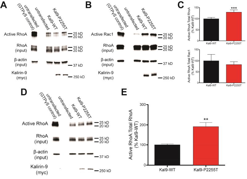
Background: Large-scale genetic studies have revealed that rare sequence variants, including single nucleotide variants (SNVs), in glutamatergic synaptic genes are enriched in schizophrenia patients. However, the majority are too rare to show any association with disease and have not been examined functionally. One such SNV, KALRN-P2255T, displays a penetrance that greatly exceeds that of previously identified schizophrenia-associated SNVs. Therefore, we sought to characterize its effects on the function of kalirin (Kal)-9, a dual Ras-related C3 botulinum toxin substrate 1 and Ras homologue gene family, member A (RhoA) guanine nucleotide exchange factor, upregulated in human schizophrenia brain tissue.

Methods: Kal9 was overexpressed in primary rat cortical neurons or human embryonic kidney 293 (HEK293) cells. The effects of the P2255T variant on dendritic branching, dendritic spine morphology, protein and messenger RNA stability, and catalytic activity were examined.

Results: Kal9-P2255T leads to diminished basal dendritic branching and dendritic spine size, compared with wild-type Kal9. The P2255T SNV directly affected Kal9 protein function, causing increased RhoA activation in HEK293 cells, but had no effect on Ras-related C3 botulinum toxin substrate 1 activation. Consistent with human postmortem findings, we found that Kal9-P2255T protein levels were higher than those of wild-type Kal9 in neurons. Increased messenger RNA stability was detected in HEK293 cells, indicating that this was the cause of the higher protein levels. When analyzed together, increased intrinsic RhoA guanine nucleotide exchange factor catalytic activity combined with increased messenger RNA expression led to net enhancement of RhoA activation, known to negatively impact neuronal morphology.

Conclusions: Taken together, our data reveal a novel mechanism for disease-associated SNVs and provide a platform for modeling morphological changes in mental disorders.

Keywords: Dendrites; Dendritic spines; Guanine nucleotide exchange factors; Kalirin; Schizophrenia; Single nucleotide variants.


Conflict of interest statement

Conflicts of interest

The authors report no biomedical financial interests or potential conflicts of interest.

Figures

Figure 1
Figure 1
KALRN associations with SZ and expression in mature neurons. A. Schematic of the Kal7 and Kal9 proteins, showing catalytic domains, structural motifs, and upstream and downstream signaling pathways. The location of the P2255T substitution in Kal9 is indicated by an arrow. Asterisk indicates the PDZ-binding domain specific to Kal7. The location of the P2255T substitution is shown. B. Hypothetical KALRN interaction network, predicted based on co-expression, pathway, and physical interaction, and weighted with regard to cellular component gene ontology. Genes shown in yellow are also among those residing in SZ-associated loci identified by a large-scale GWAS. A high-resolution rendering of this network is included in Supplemental Figure S1. C. Hypergeometric testing reveals a significant overlap between those genes in the hypothetical KALRN network and those identified by GWAS. D. SIM imaging of mature (DIV 25–26) cortical neurons demonstrates that endogenous kalirin-7 is localized in the majority of PSD95-containing spines, whereas kalirin-9 is largely excluded from spines. E. Proportions of spines with Kal7 and Kal9 expression in distinct nanodomains. Blue: no expression; red: head only; green: head and neck; purple: head and base: light blue: neck only; orange: base only. F. Spines lacking kalirin-7 are significantly smaller than those with kalirin-7 (N=98 spines). The presence of kalirin-9 in a spine has no bearing on its size (N=113 spines). Cells from three independent experiments were analyzed. Data are mean ± SEM. *, p<0.05.
Figure 2
Figure 2
Kal9-P2255T diminishes basal dendrite branching in cortical pyramidal neurons. A. DIV25–26 neurons transfected with either GFP alone, or with GFP and either Kal9-WT or Kal9-P2255T (upper panels); and traces used for Sholl analysis (lower panels). B. Sholl analysis reveals a significant reduction in basal dendrites of neurons expressing Kal9-P2255T extending 25 and 50 µm from the soma as compared to Kal9-WT. Apical dendrites were unchanged. N=20–35 neurons per transfection condition from three independent experiments. Data are mean ± SEM. **, p<0.01; ***, p<0.005 (Kal9-WT vs. Kal9-P2255T). †, p<0.05; ††, p<0.01; †††, p<0.005 (Kal9-WT vs. GFP). ‡, p<0.05; ‡‡, p<0.01; ‡‡‡, p<0.005 (Kal9-P2255T vs. GFP).
Figure 3
Figure 3
Kal9-P2255T fails to induce the increases in dendritic spine dimensions seen following Kal9-WT overexpression. A. DIV25–26 cortical pyramidal neurons transfected with either GFP alone, or with GFP and either Kal9-WT or Kal9-P2255T. B. Area and breadth of dendritic spine heads are significantly reduced in neurons expressing Kal9-P2255T as compared to Kal9-WT. Spine length and density were unaffected. 11–18 neurons per transfection condition from three independent experiments were analyzed. Data are mean ± SEM. **, p<0.01; ***, p<0.005.
Figure 4
Figure 4
Kal9-P2255T protein and mRNA are expressed more highly than Kal9-WT. A. DIV 28 cortical pyramidal neurons transfected with GFP and either Kal9-WT or Kal9-P2255T and stained for GFP and c-Myc. B, C. Fluorescence intensity of Kal9-P2255T is greater than that of Kal9-WT at both 72 h (B) and 14 d (C) post-transfection. D. The ratio of c-Myc to GFP intensity reveals that the difference in expression at 72 h is not due to a difference in transfection efficiency. N=68–131 neurons per transfection condition from three independent experiments. E. Isolated RNA from hEK293 cells transfected with either Kal9-WT or Kal9-P2255T was subjected to qRT-PCR. Following normalization to β-actin expression, Kal9-P2255T mRNA levels are shown to be increased compared to Kal9-WT. Data are from four independent experiments. F. Kal9-WT and Kal9-P2255T transfected hEK293 cells treated with ActD for 4, 8, or 12 hours show differential rates of Kal9 transcript degradation. Half-life was calculated as ln(2)/-slope. t1/2 = 6.2 hours for Kal9-WT and 15.3 hours for Kal9-P2255T. Dashed lines represent 95% confidence intervals. Data are from three independent experiments. G. mfold software predicts the disruption of an interior loop in the secondary structure of Kal9-P2255T mRNA. The nucleotide coding for amino acid 2255 resides in within the circles overlaying the images. H. Western blots of lysates from hEK293 cells transfected with GFP alone, or GFP and either Kal9-WT or Kal9-P2255T. I. When normalized to β-tubulin, Kal9-P2255T is shown to be expressed more highly than Kal9-WT. J. CHX treatment of transfected hEK293 cells demonstrates that there is no change in the stability of Kal9-P2255T versus Kal9-WT protein (see Supplemental Figure S2 for blot images). Data are from three independent experiments. All data are mean ± SEM. *, p<0.05; **, p<0.01; ***, p<0.005.
Figure 5
Figure 5
The P2255T substitution increases Kal9 RhoA-GEF catalytic activity. A. Following transfection of hEK293 cells with amounts of plasmid to account for altered Kal9 protein levels, Western blotting reveals that Kal9-P2255T-transfected cells display higher levels of GTP-bound RhoA than Kal9-WT-transfected cells. B. Under the same transfection conditions as in A, GTP-bound Rac1 levels show no difference between the Kal9-WT and Kal9-P2255T. C. Quantification of the blots in A (upper panel) and B (lower panel). Blots were normalized to total RhoA or Rac1 levels, and β-actin levels. Data are from 4–5 independent experiments. D, E. When equal amounts of Kal9-WT and Kal9-P2255T plasmids are used for transfection, an even greater level of RhoA activation is seen in the Kal9-P2255T condition than that in the experiments described in A. Data are from three independent experiments. All data are mean ± SEM. **, p<0.01; ***, p<0.005.

Comment in

References

    1. Rutkowski TP, Schroeder JP, Gafford GM, Warren ST, Weinshenker D, Caspary T, et al. Unraveling the genetic architecture of copy number variants associated with schizophrenia and other neuropsychiatric disorders. Journal of neuroscience research 2016 - PMC - PubMed
    1. Gejman PV, Sanders AR, Duan J. The role of genetics in the etiology of schizophrenia. The Psychiatric clinics of North America. 2010;33:35–66. - PMC - PubMed
    1. Fromer M, Pocklington AJ, Kavanagh DH, Williams HJ, Dwyer S, Gormley P, et al. De novo mutations in schizophrenia implicate synaptic networks. Nature. 2014;506:179–184. - PMC - PubMed
    1. Hall J, Trent S, Thomas KL, O'Donovan MC, Owen MJ. Genetic risk for schizophrenia: convergence on synaptic pathways involved in plasticity. Biol Psychiatry. 2015;77:52–58. - PubMed
    1. Purcell SM, Moran JL, Fromer M, Ruderfer D, Solovieff N, Roussos P, et al. A polygenic burden of rare disruptive mutations in schizophrenia. Nature. 2014;506:185–190. - PMC - PubMed

Publication types

MeSH terms

Substances