

SCARF-1 promotes adhesion of CD4+ T cells to human hepatic sinusoidal endothelium under conditions of shear stress
- PMID: 29242513
- PMCID: PMC5730566
- DOI: 10.1038/s41598-017-17928-4
SCARF-1 promotes adhesion of CD4+ T cells to human hepatic sinusoidal endothelium under conditions of shear stress
Abstract
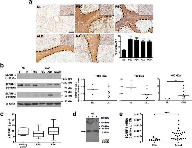
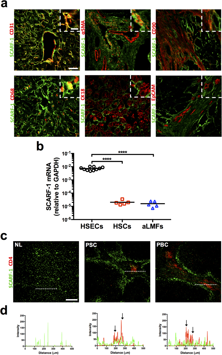
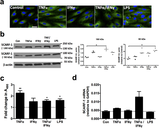
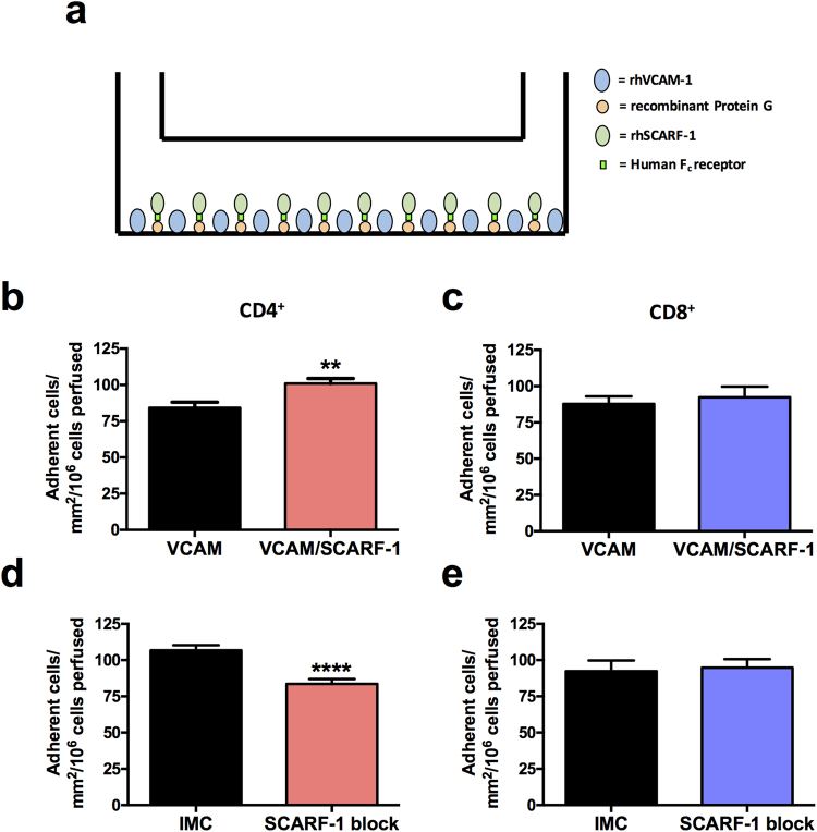
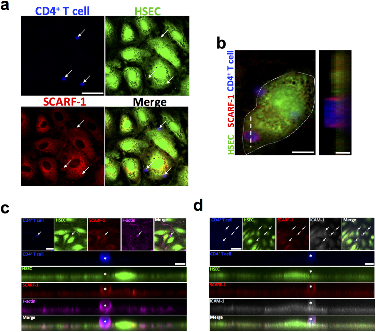
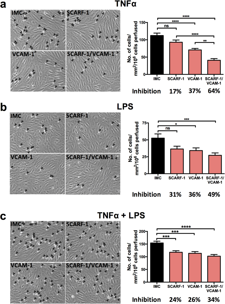
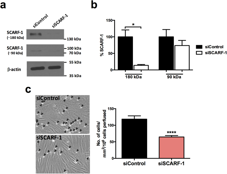
Liver-resident cells are constantly exposed to gut-derived antigens via portal blood and, as a consequence, they express a unique repertoire of scavenger receptors. Whilst there is increasing evidence that the gut contributes to chronic inflammatory liver disease, the role of scavenger receptors in regulating liver inflammation remains limited. Here, we describe for the first time the expression of scavenger receptor class F, member 1 (SCARF-1) on hepatic sinusoidal endothelial cells (HSEC). We report that SCARF-1 shows a highly localised expression pattern and co-localised with endothelial markers on sinusoidal endothelium. Analysis of chronically inflamed liver tissue demonstrated accumulation of SCARF-1 at sites of CD4+ T cell aggregation. We then studied the regulation and functional role of SCARF-1 in HSEC and showed that SCARF-1 expression by HSEC is regulated by proinflammatory cytokines and bacterial lipopolysaccharide (LPS). Furthermore, SCARF-1 expression by HSEC, induced by proinflammatory and gut-derived factors acts as a novel adhesion molecule, present in adhesive cup structures, that specifically supports CD4+ T cells under conditions of physiological shear stress. In conclusion, we show that SCARF-1 contributes to lymphocyte subset adhesion to primary human HSEC and could play an important role in regulating the inflammatory response during chronic liver disease.
Conflict of interest statement
The authors declare that they have no competing interests.
Figures
Similar articles
-
Vascular adhesion protein-1 mediates adhesion and transmigration of lymphocytes on human hepatic endothelial cells.J Immunol. 2002 Jul 15;169(2):983-92. doi: 10.4049/jimmunol.169.2.983. J Immunol. 2002. PMID: 12097405
-
Common lymphatic endothelial and vascular endothelial receptor-1 mediates the transmigration of regulatory T cells across human hepatic sinusoidal endothelium.J Immunol. 2011 Apr 1;186(7):4147-55. doi: 10.4049/jimmunol.1002961. Epub 2011 Mar 2. J Immunol. 2011. PMID: 21368224 Free PMC article.
-
CD151 supports VCAM-1-mediated lymphocyte adhesion to liver endothelium and is upregulated in chronic liver disease and hepatocellular carcinoma.Am J Physiol Gastrointest Liver Physiol. 2017 Aug 1;313(2):G138-G149. doi: 10.1152/ajpgi.00411.2016. Epub 2017 May 4. Am J Physiol Gastrointest Liver Physiol. 2017. PMID: 28473332 Free PMC article.
-
Cooperation of liver cells in health and disease.Adv Anat Embryol Cell Biol. 2001;161:III-XIII, 1-151. doi: 10.1007/978-3-642-56553-3. Adv Anat Embryol Cell Biol. 2001. PMID: 11729749 Review.
-
Human hepatic sinusoidal endothelial cells can be distinguished by expression of phenotypic markers related to their specialised functions in vivo.World J Gastroenterol. 2006 Sep 14;12(34):5429-39. doi: 10.3748/wjg.v12.i34.5429. World J Gastroenterol. 2006. PMID: 17006978 Free PMC article. Review.
Cited by
-
Considerations for feature selection using gene pairs and applications in large-scale dataset integration, novel oncogene discovery, and interpretable cancer screening.BMC Med Genomics. 2020 Oct 22;13(Suppl 10):148. doi: 10.1186/s12920-020-00778-x. BMC Med Genomics. 2020. PMID: 33087122 Free PMC article.
-
Responsible Genes for Neuronal Migration in the Chromosome 17p13.3: Beyond Pafah1b1(Lis1), Crk and Ywhae(14-3-3ε).Brain Sci. 2021 Dec 30;12(1):56. doi: 10.3390/brainsci12010056. Brain Sci. 2021. PMID: 35053800 Free PMC article. Review.
-
Protocol to study monocyte transmigration across primary human liver endothelial cells under physiological shear flow conditions in vitro.STAR Protoc. 2024 Dec 20;5(4):103431. doi: 10.1016/j.xpro.2024.103431. Epub 2024 Nov 5. STAR Protoc. 2024. PMID: 39504247 Free PMC article.
-
Dietary antarctic krill improves antioxidant capacity, immunity and reduces lipid accumulation, insights from physiological and transcriptomic analysis of Plectropomus leopardus.BMC Genomics. 2024 Feb 26;25(1):210. doi: 10.1186/s12864-024-10099-3. BMC Genomics. 2024. PMID: 38408914 Free PMC article.
-
Prognostic Value and Potential Immunoregulatory Role of SCARF1 in Hepatocellular Carcinoma.Front Oncol. 2020 Sep 29;10:565950. doi: 10.3389/fonc.2020.565950. eCollection 2020. Front Oncol. 2020. PMID: 34354939 Free PMC article.
References
Publication types
MeSH terms
Substances
Grants and funding
- MR/M009157/1/MRC_/Medical Research Council/United Kingdom
- 097162/Z/11/Z/WT_/Wellcome Trust/United Kingdom
- G0300101/MRC_/Medical Research Council/United Kingdom
- G0300102/MRC_/Medical Research Council/United Kingdom
- G0700301/MRC_/Medical Research Council/United Kingdom
- G0802577/MRC_/Medical Research Council/United Kingdom
- WT_/Wellcome Trust/United Kingdom
- BB/N018869/1/BB_/Biotechnology and Biological Sciences Research Council/United Kingdom
- MC_PC_14123/MRC_/Medical Research Council/United Kingdom
- G0400496/MRC_/Medical Research Council/United Kingdom
- CRUK_/Cancer Research UK/United Kingdom
LinkOut - more resources
Full Text Sources
Other Literature Sources
Research Materials
