. 2017 Dec 14;7(1):17565.
doi: 10.1038/s41598-017-17770-8.

The celecoxib derivatives AR-12 and AR-14 induce autophagy and clear prion-infected cells from prions

Affiliations

The celecoxib derivatives AR-12 and AR-14 induce autophagy and clear prion-infected cells from prions

Basant A Abdulrahman et al. Sci Rep. .

Abstract

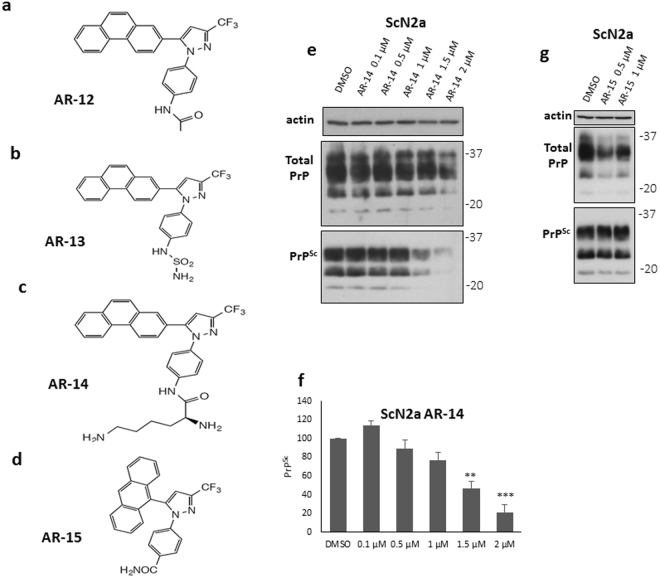
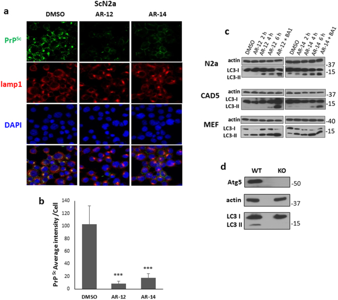
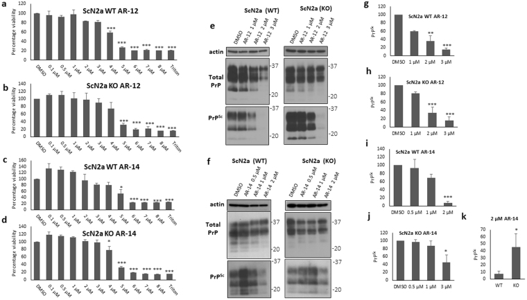
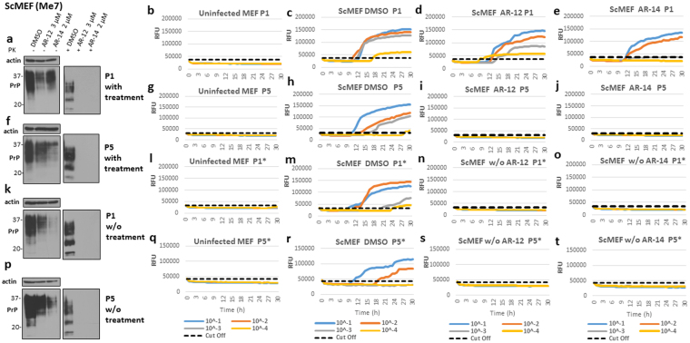
Prion diseases are fatal infectious neurodegenerative disorders that affect both humans and animals. The autocatalytic conversion of the cellular prion protein (PrPC) into the pathologic isoform PrPSc is a key feature in prion pathogenesis. AR-12 is an IND-approved derivative of celecoxib that demonstrated preclinical activity against several microbial diseases. Recently, AR-12 has been shown to facilitate clearance of misfolded proteins. The latter proposes AR-12 to be a potential therapeutic agent for neurodegenerative disorders. In this study, we investigated the role of AR-12 and its derivatives in controlling prion infection. We tested AR-12 in prion infected neuronal and non-neuronal cell lines. Immunoblotting and confocal microscopy results showed that AR-12 and its analogue AR-14 reduced PrPSc levels after only 72 hours of treatment. Furthermore, infected cells were cured of PrPSc after exposure of AR-12 or AR-14 for only two weeks. We partially attribute the influence of the AR compounds on prion propagation to autophagy stimulation, in line with our previous findings that drug-induced stimulation of autophagy has anti-prion effects in vitro and in vivo. Taken together, this study demonstrates that AR-12 and the AR-14 analogue are potential new therapeutic agents for prion diseases and possibly protein misfolding disorders involving prion-like mechanisms.


Conflict of interest statement

The authors declare that they have no competing interests.

Figures

Figure 1
Figure 1
Short-term treatment with AR-12 reduces PrPSc levels in three persistently prion-infected cell lines. (a) Persistently prion-infected neuroblastoma cells (ScN2a; prion strain 22 L) were treated for 72 h with 0.5, 1, 1.5, 2, 2.5 or 3 µM of AR-12. Solvent only-treated cells (DMSO) were used as control. Cells were lysed and lysates split into two halves. One was treated with proteinase K (PK; 20 µg/ml for 30 min at 37 °C) and both subjected to immunoblot analysis. Immunoblot was developed with anti-PrP monoclonal antibody (mAb) 4H11 and the blot was re-probed with a mAb for actin (gel loading control). (b) Densitometric analysis of ScN2a immunoblots. Data are represented as a percentage of vehicle-treated (DMSO) cells from at least three independent experiments. (c) Persistently prion-infected neuronal ScCAD5 cells (prion strain 22 L) were treated for 72 h with 1, 2, 3, 4 or 5 µM of AR-12. Solvent only-treated cells (DMSO) were used as control. Immunoblot was developed with anti-PrP mAb 4H11 and the blot was re-probed for actin (gel loading). PrPSc was reduced after AR-12 treatment. (d) Densitometric analysis for ScCAD5 immunoblots. Data are represented as a percentage of vehicle-treated (DMSO) cells. (e) Persistently prion-infected mouse embryonic fibroblasts (MEF) (prion strain 22 L) were treated for 72 h with 0.5, 1,1.5, 2, 2.5 or 3 µM of AR-12 and analyzed as above. (f) Densitometric analysis for ScMEF immunoblots. Data are represented as a percentage of vehicle-treated (DMSO) cells. (*p < 0.05), (**p < 0.01) (***p < 0.001) considered significant.
Figure 2
Figure 2
Anti-prion effect of AR-12 analogues. (ad) Chemical structures of AR-12, AR-13, AR-14 and AR-15, respectively. (e) ScN2a cells were treated for 72 h with various concentrations of AR-14 (0.1, 0.5, 1, 1.5 and 2 µM). Solvent only-treated cells (DMSO) were used as control. Immunoblot was developed with anti-PrP mAb 4H11 and re-probed for actin. (f) Densitometric analysis for ScN2a immunoblots. Data are represented as a percentage of vehicle-treated (DMSO) cells. (g) ScN2a cells were treated for 72 h with 0.5 and 1 µM of AR-15. Solvent only-treated cells (DMSO) were used as control. Immunoblot was developed with anti-PrP mAb 4H11 and re-probed for actin. (**p < 0.01), (***p < 0.001) considered significant.
Figure 3
Figure 3
Immunofluorescence analysis of AR-12 and AR-14 effects and induction of autophagy. (a) ScN2a cells were treated with AR-12 or AR-14 for 72 h. DMSO-treated cells were used as a control. Cells were fixed and confocal microscopy staining for PrPSc (mAb 4H11, green) and lamp1 (red) was done. Nuclei were stained with DAPI (blue). Lower panel shows merge. (b) The overall immunofluorescence intensity of PrPSc of five images for either DMSO, AR-12 or AR-14 treated cells was measured. Overall intensity was divided by the cell number contained from the same image quantified by ImageJ “analyze particle” command to calculate the averaged immunofluorescence intensity per cell. (c) N2a, CAD5 and MEF cells were treated with AR-12 (3 µM) or AR-14 (2 µM), respectively, for 2, 4 and 6 h, or AR compound plus Bafilomycin A1 (BA1) (100 nm, for 4 h). Solvent only-treated cells (DMSO) were used as control. Immunoblots were developed with anti-LC3 mAb (autophagy marker) and mAb for actin (gel loading control). Treatment both with AR-12 and AR-14 showed a time dependent increase in LC3-II levels. BA1 treated cells had the highest expression level of LC3-II due to the block on autophagic flux and lysosomal function. (d) N2a cells were established with knock-out in the autophagy gene ATG5. Immunoblot compares N2a knockout (KO) to wild-type (WT) cells. Immunoblot was probed for Atg5, LC3-I/II and actin. There are no Atg5 and LC3-II bands in N2a-KO cells.
Figure 4
Figure 4
AR-12 and AR-14 anti-prion effects involve autophagy. (a–d) XTT viability assay of ScN2a WT and ScN2a Atg5 KO cells treated for 72 h with AR-12 or AR-14. (e,f) Persistently prion-infected WT (left side) and KO (right side) ScN2a cells (strain 22 L) were treated for 72 h with different concentrations of AR-12 and AR-14, respectively. Solvent only-treated cells (DMSO) were used as control. Immunoblots were developed with anti-PrP mAb 4H11 and re-probed for actin. (g–k) Densitometric analysis for panel e and f immunoblots, data were represented as a percentage of vehicle (DMSO) treated cells. (*p < 0.05), (***P < 0.001) considered significant.
Figure 5
Figure 5
AR-12 long-term treatment cured prion infected ScN2a cells (22 L). ScN2a cells were treated with AR-12 (3 µM) or AR-14 (2 µM). DMSO treatment was used as control. Treatment was continued for five passages (20 days). Then, the treatment was stopped and cells were passaged five times again (20 days). (a,f,k and p) Immunoblots showing the effect of treatment with AR compounds on PrPSc throughout the experiment compared to DMSO treated cells. Immunoblots were developed with anti-PrP mAb 4H11 and probed for actin. (b,g,l and q) RT-QuIC analysis for uninfected N2a cells at every passage. Recombinant mouse PrP was used as substrate. Each quadruplicate RT-QuIC reaction was seeded with 2 μl of cell lysate (at dilutions 10−1 to 10−4). The average increase of Thioflavin-T fluorescence of replicate wells is plotted as a function of time. Y-axis represents relative fluorescent units (RFU) and x-axis time in hours. (c,h,m and r) RT-QuIC analysis for DMSO-treated cells. Passages 1 and 5 (P1 and P5) are shown (c,h). After discontinuation of the treatment, passages 1 and 5 (P1* and P5*) are shown (m,r). (d,i,n and s) RT-QuIC-analysis for cells treated with AR-12 (3 µM) for 20 days. Passages 1 and 5 (P1 and P5) are shown (d,i). After discontinuation of the treatment, passages 1 and 5 (P1* and P5*) are shown (n,s). (e,j,o and t) RT-QuIC analysis for cells treated with AR-14 (2 µM) for 20 days. Passages 1 and 5 (P1 and P5) are shown (e,j). After discontinuation of the treatment, passages 1 and 5 (P1* and P5*) are shown (o,t).
Figure 6
Figure 6
AR-12 and AR-14 long-term treatment cured prion infection in ScMEF cells infected with Me7 prions. (a–d) ScMEFs (Me7) were treated with AR-12 (3 µM) or AR-14 (2 µM). DMSO-treated cells were used as a control. Treatment was continued for five passages (20 days). Then, the treatment was stopped and cells were passaged five times again (20 days). (a,f,k and p) Immunoblots showing the effect of treatment with AR compounds on PrPSc throughout the experiment compared to DMSO-treated cells. Immunoblots were developed with anti-PrP mAb 4H11 and re-probed for actin. (b,g,l and q) RT-QuIC analysis for uninfected MEF cells at every passage. Each quadruplicate RT-QuIC reaction was seeded with 2 μl of cell lysate (at dilutions 10−1 to 10−4). The average increase of Thioflavin-T fluorescence of replicate wells is plotted as a function of time. Y-axis represents relative fluorescent units (RFU) and x-axis time in hours. (c,h,m and r) RT-QuIC analysis for DMSO-treated cells. Passages 1 and 5 (P1 and P5) are shown (c,h). After discontinuation of the treatment, passages 1 and 5 (P1* and P5*) are shown (m,r). (d,i,n and s) RT-QuIC-analysis for cells treated with AR-12. (e,j,o and t) RT-QuIC analysis for cells treated with AR-14.

Similar articles

Cited by

References

    1. Linden R, et al. Physiology of the Prion Protein. Physiol. Rev. 2008;88:673–728. doi: 10.1152/physrev.00007.2007. - DOI - PubMed
    1. Martins VR, et al. Prion protein: orchestrating neurotrophic activities. Curr. Issues Mol. Biol. 2010;12:63–86. - PubMed
    1. Watts JC, Westaway D. The prion protein family: Diversity, rivalry, and dysfunction. Biochim. Biophys. Acta - Mol. Basis Dis. 2007;1772:654–672. doi: 10.1016/j.bbadis.2007.05.001. - DOI - PubMed
    1. Martin-Lannerée S, et al. The Cellular Prion Protein Controls Notch Signaling in Neural Stem/Progenitor Cells. Stem Cells. 2017;35:754–765. doi: 10.1002/stem.2501. - DOI - PubMed
    1. Prusiner SB. Novel proteinaceous infectious particles cause scrapie. Science. 1982;216:136–44. doi: 10.1126/science.6801762. - DOI - PubMed

Publication types