. 2017 Dec 14;8(1):2123.
doi: 10.1038/s41467-017-02217-5.

Nuclear lamin A/C harnesses the perinuclear apical actin cables to protect nuclear morphology

Affiliations

Nuclear lamin A/C harnesses the perinuclear apical actin cables to protect nuclear morphology

Jeong-Ki Kim et al. Nat Commun. .

Erratum in

Abstract

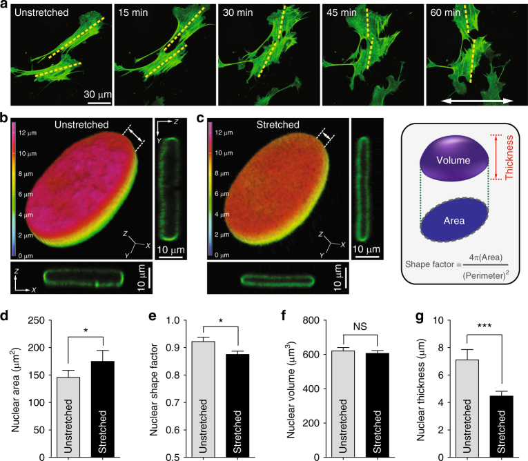
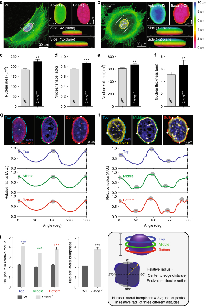
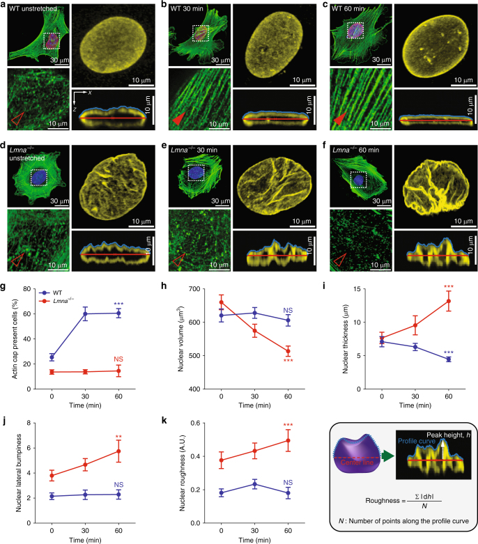
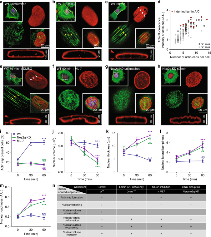
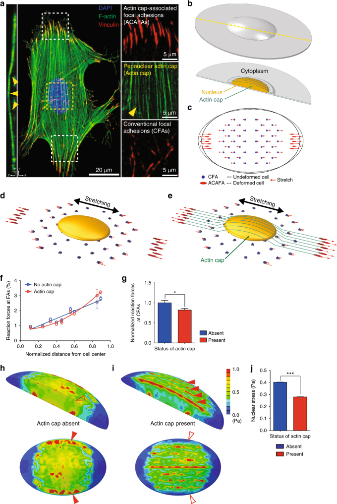
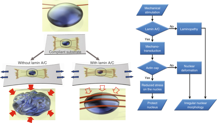
The distinct spatial architecture of the apical actin cables (or actin cap) facilitates rapid biophysical signaling between extracellular mechanical stimuli and intracellular responses, including nuclear shaping, cytoskeletal remodeling, and the mechanotransduction of external forces into biochemical signals. These functions are abrogated in lamin A/C-deficient mouse embryonic fibroblasts that recapitulate the defective nuclear organization of laminopathies, featuring disruption of the actin cap. However, how nuclear lamin A/C mediates the ability of the actin cap to regulate nuclear morphology remains unclear. Here, we show that lamin A/C expressing cells can form an actin cap to resist nuclear deformation in response to physiological mechanical stresses. This study reveals how the nuclear lamin A/C-mediated formation of the perinuclear apical actin cables protects the nuclear structural integrity from extracellular physical disturbances. Our findings highlight the role of the physical interactions between the cytoskeletal network and the nucleus in cellular mechanical homeostasis.


Conflict of interest statement

The authors declare no competing financial interests.

Figures

Fig. 1
Fig. 1
Stretch-induced nuclear morphological changes in mouse embryonic fibroblasts. a Cell re-orientation under uniaxial cyclic stretching of the substrate. Randomly seeded EGFP–LifeAct-transfected mouse embryonic fibroblasts (MEFs) onto the stretchable PDMS thin film were re-oriented in response to the substrate stretching (8% strain ratio and 1 Hz frequency) by the reorganization of actin cytoskeleton perpendicular to the substrate stretching direction (indicated by white arrow). Yellow dashed lines indicate the main axis of the actin cytoskeleton traversing the cell body. Refer to Supplementary Figure 1 for the verification of experimental setup. b, c Stretch-induced nuclear flattening. A GFP–lamin-A-transfected MEF was imaged under the same stretching condition for 1 h. Pseudo-colored z-depth rendering and xz and yz cross-sectional views of the three-dimensionally reconstructed nuclei represent stretch-induced nuclear flattening. dg Quantification of nuclear morphological changes. Footprint size (i.e., area) and shape factor estimated from a projected nuclear morphology onto the XY-plane depict a slight enlargement and elongation of the stretched nuclei (d, e). Nuclear volume and thickness (i.e., the maximum length from the basal surface to the apical surface) of the three-dimensionally reconstructed nuclei reveal that substrate stretching generates a pressing force vertical to the nucleus of adherent cells, whereas nuclear volume was conserved (f, g). To avoid out-of-focus images and to prevent photobleaching of transfected cells, each image was captured every 15 min for ~1 h (a). Overall, >30 cells were examined per condition (dg), where error bars indicate S.E.M. and statistical differences were calculated using the unpaired t-test, ***p < 0.0001, *p < 0.01, NS not significant (p > 0.05)
Fig. 2
Fig. 2
Effect of nuclear lamin A/C on nuclear morphology. af Lamin A/C-dependent difference of nuclear morphology in MEFs placed in a glass bottom dish. Representative cell and nuclear morphology in lamin A/C-present control wild-type (WT) and lamin A/C-deficient Lmna knockout (Lmna−/−) MEFs were captured by 3D reconstruction of immunofluorescence confocal images along the z-axis (a, b). Note that apical, basal, and side views of pseudo-colored 3D z-depth rendered nuclei display the differences in the nuclear surface textures. Lmna−/− cells displayed more spread (c), rounder (d), larger (e), and thicker (f) nuclei than the WT control cells. gj Comparison of lamin A/C-dependent lateral deformation of the nucleus. Tracking surface contours in three different altitudes in a nucleus displays significantly more peak positions (gray dots) in Lmna−/− cells than WT cells (gi), representing more protrusions on the nuclear surface of the Lmna−/− MEFs. Relative radius is the ratio between the nuclear center-to-edge distance and the equivalent circular radius, where top (blue), middle (green), and bottom (red) parts correspond to the top 25%, top 50%, and top 75% positions along the z-axis of the nucleus, respectively. Average number of peaks in relative radii of three different altitudes was termed by the nuclear lateral bumpiness (j). In cj, >30 cells were analyzed per condition. Error bars represent S.E.M. of averaged values and statistical differences were calculated using the unpaired t-test; ***p < 0.0001, **p < 0.001
Fig. 3
Fig. 3
Lamin A/C-dependent differential formation of an actin cap and nuclear deformation in response to substrate stretching. af Representative F-actin organization and nuclear morphology of lamin A/C-present WT (ac) and lamin A/C-deficient Lmna−/− (df) MEFs at different time points of the substrate stretching (0, 30, 60 min). Insets display details of F-actin organization in the apical region of the nucleus. Full and empty arrowheads indicate the presence and absence of the perinuclear actin cap, respectively. Nuclear morphology of lamin B1-stained nuclei (yellow) indicates distinct evolution of 3D-nuclear shape in response to substrate stretching, where maximum intensity projection onto the XY-plane was performed using upper hemispheres of the 3D-reconstructed nuclei to highlight the detailed nuclear surface texture. The cross-sectional side view was captured along the XZ-plane crossing the center of the nucleus. gk Stretch-dependent formation of an actin cap (g) and changes of nuclear volume (h), thickness (i), lateral bumpiness (j), and surface roughness (k) in WT (blue) and Lmna−/− (red) MEFs. Note that nuclear flattening with volume conversation (h, i) and nuclear surface roughening with volume reduction (j, k) are the stretch-induced characteristic features of the actin cap forming WT and actin cap non-forming Lmna−/− cells (g). In i and k, nuclear thickness was defined as the maximum height in the vertically cross-sectioned image of the 3D-reconstructed lamin B1-stained nucleus and the height variation along the apical surface was termed as surface roughness. In g, >150 cells were examined per condition and in hk, >30 lamin B1-stained nuclei were analyzed per condition. Error bars represent the S.E.M. of averaged values. Unpaired t-test was applied to compare unstretched control cells (0 min) and fully stretched cells (60 min). ***p < 0.0001, **p < 0.001, NS not significant (p > 0.05)
Fig. 4
Fig. 4
Underlying molecular mechanism of nuclear deformation. ad Substrate stretching induced formation of an actin cap and reorganization of the nuclear lamin A/C. Although unstretched MEFs placed on the compliant PDMS film typically displayed a dismantled actin cap and vertically isotropic organization of the lamin A/C (red) wrapping the thick nucleus (a), continuous substrate stretching induces the formation of apical actin stress fibers (>30 min, b, c) and the nucleus became flattened (60 min, c). Note that the prolonged stretching time induces thickening of the stress fibers in the actin cap (bd) and the formation of indented marks on the lamin A/C-stained apical surface of the nucleus along the actin cap (c, d), where lamin A/C forms an apically polarized dorm structure (b vs. c). Indented marks on lamin A/C were detected only after 60 min of stretching (c, d). eh Two molecular mechanisms of stretch-induced nuclear morphological responses: actomyosin contractility and nucleus-cytoskeletal connectivity. Although lamin A/C was present, treatment of a myosin light-chain kinase (MLCK) inhibiting drug, ML-7 (e vs. f) and depletion of nesprin-2G (Nes2g) proteins (g vs. h) abrogated the stretching induced formation of an actin cap, where severe nuclear deformation was detected in response to substrate stretching (f, h). im Quantification of the actin cap formation and nuclear morphology of ML-7 treated (purple) and nesprin-2G depleted (green) MEFs in response to the substrate stretching. Note that the loss of actomyosin contractility or nucleus-cytoskeletal connectivity did not induce the formation of an actin cap (i) but reduced the nuclear volume (j), where severe nuclear morphological deformation was detected (km) in response to substrate stretching. n Summary of the relationship between the actin cap formation and nuclear morphological responses. In i, >150 cells were examined per condition and in jm, >30 lamin B1-stained nuclei were analyzed per condition. Error bars represent S.E.M. of averaged values and statistical differences were calculated by unpaired t-test between unstretched control cells (0 min) and fully stretched cells (60 min). ***p < 0.0001, **p < 0.001, NS not significant (p > 0.05)
Fig. 5
Fig. 5
Actin cap induced reduction of nuclear stress by focusing focal-adhesion forces on the periphery of the cell. a Organization of an actin cap and actin cap-associated focal adhesions in a MEF. Nucleus (blue), F-actin (green), and vinculin-stained focal adhesions (red) were visualized by maximum intensity projection of the z-stacked immunofluorescence confocal images. Vertical cross-section depicts the characteristic topology of the actin cap. Parallel-aligned actin cap (yellow arrowhead) are terminated by actin cap-associated focal adhesions (ACAFAs) at the basal surface of the cell, which are larger than the conventional focal adhesions (CFAs). b, c 3D finite element model of an adherent MEF. Cross-section (yellow dotted line) of continuum solid elements for cytoplasm reveals a perinuclear region, a continuum solid for nucleus, and 3D beams of the actin cap (b). Red arrows in projected bottom view correspond to the linear stretch equivalent to 8% strain (c). dg Effects of the actin cap on the forces acting on focal adhesions under stretching. Vector results of the force in the focal adhesions are depicted in the cell model in the absence (d) and presence (e) of an actin cap. Radial distribution of reaction forces at focal adhesions (f) and the average values (g) were calculated depending on the formation of an actin cap in f and g, 56 focal adhesions included in the cell model were assessed. hj Nuclear stress distribution in the stretched cell. Isometrics of von-Mises stresses in the absence (h) and presence (i) of an actin cap reveal that external forces are ~30% less transferred to the nucleus of the actin cap forming cell than the actin cap absent cell (j). In h and i, the cross-sectioned nuclei were color-coded to clearly show the stress distribution, where scale bars indicate the magnitude of mises stresses. In j, to facilitate quantitative comparison, nuclear stress was expressed as von-Mises stress that represent a non-directional scalar quantity. Values were extracted from 19,248 different nodes inside the nucleus. In f, g, and j statistical analyses were performed using the unpaired t-test, where error bars represent S.E.M. of averaged values; ***p < 0.0001, *p < 0.05
Fig. 6
Fig. 6
Summary of the functional interplay between lamin A/C and actin cap in response to the external physical stimuli. A schematic flow chart of the lamin A/C-mediated cellular mechanotransduction and the role of the actin cap in protecting nuclear morphology. The external mechanical stimuli trigger the formation of actin cap in lamin A/C-present cells, which reduces the stress transferred to the nucleus and maintains the nuclear morphology. Laminopathic cells featuring the loss of the actin cap typically display the irregular nuclear morphology

Similar articles

Cited by

References

    1. Kudlow BA, Kennedy BK, Monnat RJ. Werner and Hutchinson–Gilford progeria syndromes: mechanistic basis of human progeroid diseases. Nat. Rev. Mol. Cell Biol. 2007;8:394–404. doi: 10.1038/nrm2161. - DOI - PubMed
    1. Quijano-Roy S, et al. De novo LMNA mutations cause a new form of congenital muscular dystrophy. Ann. Neurol. 2008;64:177–186. doi: 10.1002/ana.21417. - DOI - PubMed
    1. Fatkin D, et al. Missense mutations in the rod domain of the lamin A/C gene as causes of dilated cardiomyopathy and conduction-system disease. N. Engl. J. Med. 1999;341:1715–1724. doi: 10.1056/NEJM199912023412302. - DOI - PubMed
    1. Moulson CL, et al. Homozygous and compound heterozygous mutations in ZMPSTE24 cause the laminopathy restrictive dermopathy. J. Invest. Dermatol. 2005;125:913–919. doi: 10.1111/j.0022-202X.2005.23846.x. - DOI - PMC - PubMed
    1. Shackleton S, et al. LMNA, encoding lamin A/C, is mutated in partial lipodystrophy. Nat. Genet. 2000;24:153–156. doi: 10.1038/72807. - DOI - PubMed

Publication types

LinkOut - more resources