. 2017 Dec 14;7(1):17563.
doi: 10.1038/s41598-017-17541-5.

IL-33 is induced in undifferentiated, non-dividing esophageal epithelial cells in eosinophilic esophagitis

Affiliations

IL-33 is induced in undifferentiated, non-dividing esophageal epithelial cells in eosinophilic esophagitis

J Travers et al. Sci Rep. .

Abstract

The molecular and cellular etiology of eosinophilic esophagitis (EoE), an emerging tissue-specific allergic disease, involves dysregulated gene expression in esophageal epithelial cells. Herein, we assessed the esophageal expression of IL-33, an epithelium-derived alarmin cytokine, in patients with EoE. IL-33 protein was markedly overexpressed within the nuclei of a subpopulation of basal layer esophageal epithelial cells in patients with active EoE compared to control individuals. IL-33 exhibited dynamic expression as levels normalized upon EoE remission. IL-33-positive basal epithelial cells expressed E-cadherin and the undifferentiated epithelial cell markers keratin 5 and 14 but not the differentiation marker keratin 4. Moreover, the IL-33-positive epithelial cells expressed the epithelial progenitor markers p75 and p63 and lacked the proliferation markers Ki67 and phospho-histone H3. Additionally, the IL-33-positive cells had low expression of PCNA. IL-33 expression was detected in ex vivo-cultured primary esophageal epithelial cells in a subpopulation of cells lacking expression of proliferation markers. Collectively, we report that IL-33 expression is induced in an undifferentiated, non-dividing esophageal epithelial cell population in patients with active EoE.


Conflict of interest statement

M.E.R. is a consultant for N.K.T. Therapeutics, Pulm One, Spoon Guru, Celgene, Shire, Astra Zeneca, and Novartis and has an equity interest in the first three listed and Immune Pharmaceuticals and royalties from reslizumab (Teva Pharmaceuticals). M.E.R. is an inventor of several patents, owned by Cincinnati Children’s. All of the other authors have no potential conflicts to disclose.

Figures
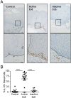
Figure 1
Figure 1
Subcellular localization of IL-33 in esophageal epithelial cells. (A,B) Immunohistochemistry for IL-33 protein expression in representative esophageal biopsies from healthy control individuals (Control, left panel), patients with active EoE (Active EoE, middle panel), or patients with inactive EoE (Inactive EoE, right panel) using mouse anti–IL-33 antibody. In (A), the bottom row is a high-power view of the area enclosed in the black square. The black dashed lines indicate the basement membrane. Scale bars are both 20 µm. Biopsies from 19 controls, 20 patients with active EoE, and 7 patients with inactive EoE were stained. (B) Quantification of the proportion of basal layer cells in each biopsy with IL-33 expression. Mean ± standard error of the mean is depicted. ****p < 0.0001.
Figure 2
Figure 2
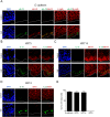
Epithelium-specific protein expression in esophageal tissue. (AD) Immunofluorescence of esophageal biopsies from control individuals (top row) or patients with active EoE (bottom row). Nuclei are indicated by DAPI staining (blue). Green and red indicate staining with the indicated antibodies. The white dashed lines indicate the basement membrane. Scale bar is 20 µm. Images are representative of biopsies from 4 or 5 patients with active EoE and 4 or 5 control individuals. (E) Quantification of the proportion of IL-33–positive basal layer cells from active EoE biopsies with strong expression of the indicated marker from (AD). Mean ± standard error of the mean is depicted. E-cadh, E-cadherin; DAPI, 4′,6-diamidino-2-phenylindole; gIL-33, goat anti–IL-33 antibody; mIL-33, mouse anti–IL-33 antibody; KRT, keratin.
Figure 3
Figure 3
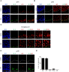
Cell cycle and differentiation status in vivo in esophageal tissue. (AD) Immunofluorescence of esophageal biopsies from control individuals (top row) or patients with active EoE (bottom row). Nuclei are indicated by DAPI staining (blue). Green and red indicate staining with the indicated antibodies. The white dashed lines indicate the basement membrane. Scale bar is 20 µm. White asterisks indicate cells with high expression of PCNA. Images are representative of biopsies from 3–6 patients with active EoE and 3–6 control individuals. (E) Quantification of the number of IL-33–positive basal layer cells from active EoE biopsies with strong expression of the indicated marker from (AD). Mean ± standard error of the mean is depicted. DAPI, 4′,6-diamidino-2-phenylindole; pH3, phospho-histone H3; PCNA, proliferating cell nuclear antigen.
Figure 4
Figure 4
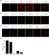
Cell cycle status of IL-33–expressing esophageal cells ex vivo. (AC) Immunofluorescence of ex vivo–cultured primary esophageal epithelial cells. Nuclei are indicated by DAPI staining (blue). Green and red indicate staining with the indicated antibodies. Images are representative of three independent experiments. (D) Quantification of the percentage of IL-33–positive primary epithelial cells with strong expression of the indicated marker. Mean ± standard error of the mean of cumulative data from three independent experiments is depicted. Scale bar is 20 µm. DAPI, 4′,6-diamidino-2-phenylindole; gIL-33, goat anti–IL-33 antibody; mIL-33, mouse anti–IL-33 antibody; KRT, keratin; pH 3, phospho-histone H3; PCNA, proliferating cell nuclear antigen.
Figure 5
Figure 5
Summary of markers of IL-33–positive esophageal epithelial cells in EoE. In the esophagus of patients with active EoE, IL-33 (yellow circle) is induced in the nuclei of interpapillary basal layer cells. In both patients with EoE and healthy individuals, these basal layer cells express E-cadherin, undifferentiated keratins (KRT5, KRT14), and epithelial progenitor markers (p75, p63). IL-33+ cells do not express the differentiation marker KRT4 or the proliferation markers Ki67, pH 3, or PCNA. Gray vertical bar indicates epithelial cell layers expressing KRT5 and KRT14, and black vertical bar indicates those layers expressing KRT4. For clarity, papillae are not depicted. E-cadh, E-cadherin; EoE, eosinophilic esophagitis; IL, interleukin; KRT, keratin; PCNA, proliferating cell nuclear antigen; pH3, phospho-histone H3.

Similar articles

Cited by

References

    1. Davis BP, Rothenberg ME. Mechanisms of Disease of Eosinophilic Esophagitis. Annu Rev Pathol. 2016;11:365–393. doi: 10.1146/annurev-pathol-012615-044241. - DOI - PMC - PubMed
    1. Travers J, Rothenberg ME. Eosinophils in mucosal immune responses. Mucosal Immunol. 2015;8:464–475. doi: 10.1038/mi.2015.2. - DOI - PMC - PubMed
    1. Liew FY, Girard JP, Turnquist HR. Interleukin-33 in health and disease. Nat Rev Immunol. 2016;16:676–689. doi: 10.1038/nri.2016.95. - DOI - PubMed
    1. Martin NT, Martin MU. Interleukin 33 is a guardian of barriers and a local alarmin. Nat Immunol. 2016;17:122–131. doi: 10.1038/ni.3370. - DOI - PubMed
    1. Bouffi C, et al. IL-33 markedly activates murine eosinophils by an NF-kappaB-dependent mechanism differentially dependent upon an IL-4-driven autoinflammatory loop. J. Immunol. 2013;191:4317–4325. doi: 10.4049/jimmunol.1301465. - DOI - PMC - PubMed

Publication types

MeSH terms