

Dual repression of endocytic players by ESCC microRNAs and the Polycomb complex regulates mouse embryonic stem cell pluripotency
- PMID: 29242593
- PMCID: PMC5730570
- DOI: 10.1038/s41598-017-17828-7
Dual repression of endocytic players by ESCC microRNAs and the Polycomb complex regulates mouse embryonic stem cell pluripotency
Abstract
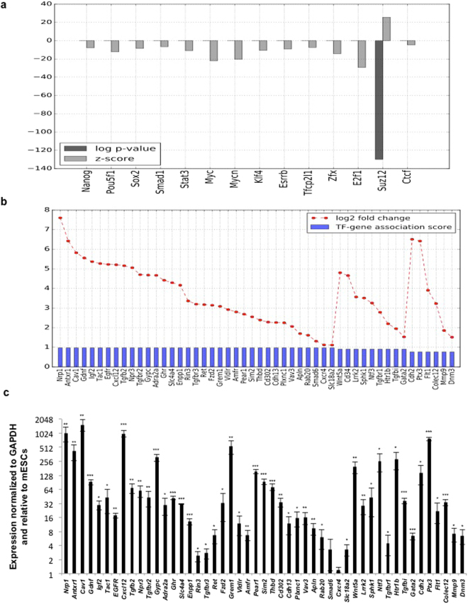
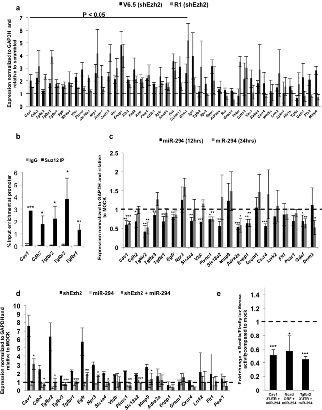
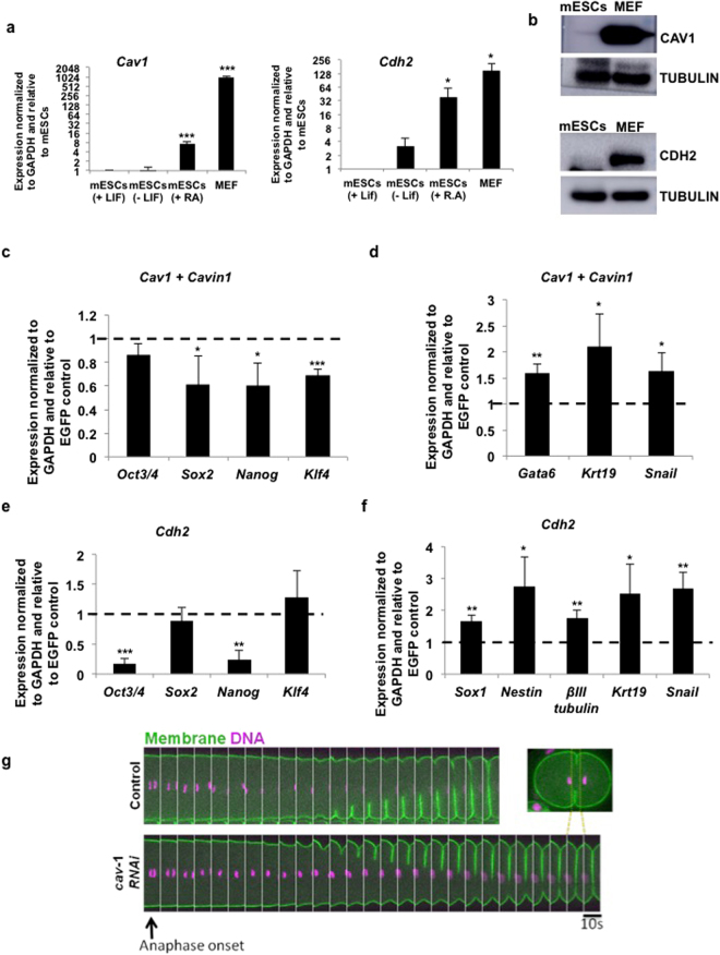
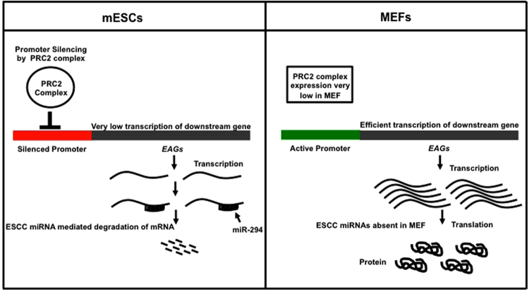
Cell fate determination in the early mammalian embryo is regulated by multiple mechanisms. Recently, genes involved in vesicular trafficking have been shown to play an important role in cell fate choice, although the regulation of their expression remains poorly understood. Here we demonstrate for the first time that multiple endocytosis associated genes (EAGs) are repressed through a novel, dual mechanism in mouse embryonic stem cells (mESCs). This involves the action of the Polycomb Repressive Complex, PRC2, as well as post-transcriptional regulation by the ESC-specific cell cycle-regulating (ESCC) family of microRNAs. This repression is relieved upon differentiation. Forced expression of EAGs in mESCs results in a decrease in pluripotency, highlighting the importance of dual repression in cell fate regulation. We propose that endocytosis is critical for cell fate choice, and dual repression may function to tightly regulate levels of endocytic genes.
Conflict of interest statement
The authors declare that they have no competing interests.
Figures
Similar articles
-
Polycomb Regulates Mesoderm Cell Fate-Specification in Embryonic Stem Cells through Activation and Repression Mechanisms.Cell Stem Cell. 2015 Sep 3;17(3):300-15. doi: 10.1016/j.stem.2015.08.009. Cell Stem Cell. 2015. PMID: 26340528
-
PRC2 specifies ectoderm lineages and maintains pluripotency in primed but not naïve ESCs.Nat Commun. 2017 Sep 22;8(1):672. doi: 10.1038/s41467-017-00668-4. Nat Commun. 2017. PMID: 28939884 Free PMC article.
-
Polycomb group RING finger proteins 3/5 activate transcription via an interaction with the pluripotency factor Tex10 in embryonic stem cells.J Biol Chem. 2017 Dec 29;292(52):21527-21537. doi: 10.1074/jbc.M117.804054. Epub 2017 Oct 20. J Biol Chem. 2017. PMID: 29054931 Free PMC article.
-
Polycomb group proteins and their roles in regulating stem cell development.Zhongguo Yi Xue Ke Xue Yuan Xue Bao. 2012 Jun;34(3):281-5. doi: 10.3881/j.issn.1000-503X.2012.03.019. Zhongguo Yi Xue Ke Xue Yuan Xue Bao. 2012. PMID: 22776663 Review.
-
The polycomb complex PRC1: composition and function in plants.J Genet Genomics. 2013 May 20;40(5):231-8. doi: 10.1016/j.jgg.2012.12.005. Epub 2013 Jan 3. J Genet Genomics. 2013. PMID: 23706298 Review.
Cited by
-
Pluripotency of embryonic stem cells lacking clathrin-mediated endocytosis cannot be rescued by restoring cellular stiffness.J Biol Chem. 2020 Dec 4;295(49):16888-16896. doi: 10.1074/jbc.AC120.014343. Epub 2020 Oct 21. J Biol Chem. 2020. PMID: 33087446 Free PMC article.
-
CLCa mediates a novel cross-talk between Wnt secretion and actin organization.Life Sci Alliance. 2025 May 2;8(7):e202402962. doi: 10.26508/lsa.202402962. Print 2025 Jul. Life Sci Alliance. 2025. PMID: 40316417 Free PMC article.
-
The Key Role of MicroRNAs in Self-Renewal and Differentiation of Embryonic Stem Cells.Int J Mol Sci. 2020 Aug 31;21(17):6285. doi: 10.3390/ijms21176285. Int J Mol Sci. 2020. PMID: 32877989 Free PMC article. Review.
-
MicroRNAs reinforce repression of PRC2 transcriptional targets independently and through a feed-forward regulatory network.Genome Res. 2019 Feb;29(2):184-192. doi: 10.1101/gr.238311.118. Epub 2019 Jan 16. Genome Res. 2019. PMID: 30651280 Free PMC article.
-
Clathrin-Mediated Endocytosis Regulates a Balance between Opposing Signals to Maintain the Pluripotent State of Embryonic Stem Cells.Stem Cell Reports. 2019 Jan 8;12(1):152-164. doi: 10.1016/j.stemcr.2018.11.018. Epub 2018 Dec 13. Stem Cell Reports. 2019. PMID: 30554918 Free PMC article.
References
-
- Li L, et al. A unique interplay between Rap1 and E-cadherin in the endocytic pathway regulates self-renewal of human embryonic stem cells. Stem Cells Dayt. Ohio. 2010;28:247–257. - PubMed
Publication types
MeSH terms
Substances
Grants and funding
LinkOut - more resources
Full Text Sources
Other Literature Sources
Research Materials
