. 2017 Dec 14;7(1):17551.
doi: 10.1038/s41598-017-17883-0.

Rheological Droplet Interface Bilayers (rheo-DIBs): Probing the Unstirred Water Layer Effect on Membrane Permeability via Spinning Disk Induced Shear Stress

Affiliations

Rheological Droplet Interface Bilayers (rheo-DIBs): Probing the Unstirred Water Layer Effect on Membrane Permeability via Spinning Disk Induced Shear Stress

Nathan E Barlow et al. Sci Rep. .

Abstract

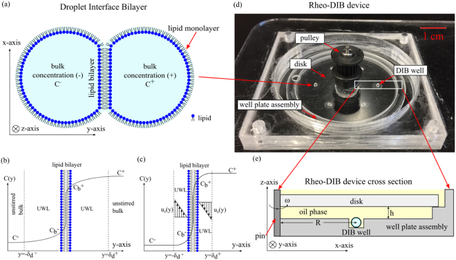
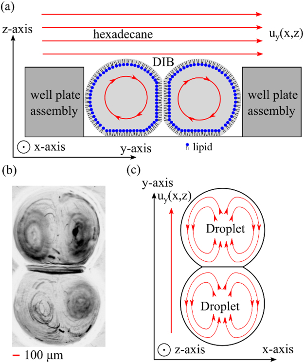
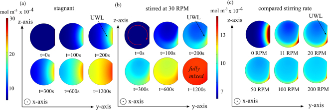
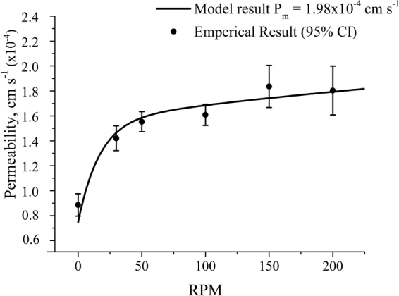
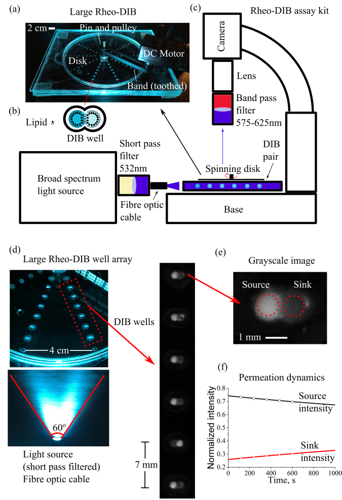
A new rheological droplet interface bilayer (rheo-DIB) device is presented as a tool to apply shear stress on biological lipid membranes. Despite their exciting potential for affecting high-throughput membrane translocation studies, permeability assays conducted using DIBs have neglected the effect of the unstirred water layer (UWL). However as demonstrated in this study, neglecting this phenomenon can cause significant underestimates in membrane permeability measurements which in turn limits their ability to predict key processes such as drug translocation rates across lipid membranes. With the use of the rheo-DIB chip, the effective bilayer permeability can be modulated by applying shear stress to the droplet interfaces, inducing flow parallel to the DIB membranes. By analysing the relation between the effective membrane permeability and the applied stress, both the intrinsic membrane permeability and UWL thickness can be determined for the first time using this model membrane approach, thereby unlocking the potential of DIBs for undertaking diffusion assays. The results are also validated with numerical simulations.


Conflict of interest statement

The authors declare that they have no competing interests.

Figures

Figure 1
Figure 1
Cartoon of droplet interface bilayer (DIB) (a) that consists of two aqueous lipid-emulsions droplets that have formed a lipid bilayer membrane at the interface. Sketch (b) of a species concentration profile across a lipid bilayer membrane (y-axis origin is on the membrane) affected by a diffusional UWL of thicknesses δd± where a solute in the bulk solution C+ on the (+) side permeates across the membrane and water layers to the (−) side bulk solution of concentration C. Note that the bilayer membrane concentration differs on either side by Cb+ and Cb. Sketch (c) of a species concentration profile across a lipid bilayer membrane with a concentration gradient on opposing sides C+ and C, where a parallel fluid flow in the bulk solution develops a laminar boundary layer at the interface. This fluid dynamic boundary layer affects the bilayer concentrations (Cb+ and Cb) and the diffusional UWL thickness δd± as a function of fluid velocity uz(y) in the z direction. Note that the thicknesses are not drawn to scale. Photograph (d) of rheo-DIB chip which can be assembled on a fluorescence microscope. A non-polar shearing media (hexadecane) is filled to 3 mm above the disk in the well plate assembly and the DIBs are formed within the DIB wells. The disk is driven by DC brushed motor and a toothed band/pulley up to 200 RPM, the maximum speed in which DIBs are stable for this configuration. Cartoon (e) of an axial-radial (x, z) cross section of rheo-DIB chip where the aqueous DIB sits in a confinement DIB well (at R = 17 mm) which is surrounded by an oil phase, and is exposed to shear stress by a disk spun about the z-axisat a determined height (h = 1 mm)above the top of the droplets and angular velocity ω.
Figure 2
Figure 2
Cartoon (a) of axial-azimuthal (y, z) cross-section with streamlines of recirculation flow in a DIB pair due to shearing flow at the top of the droplets from the non-polar phase (hexadecane) with the velocity flow profile uy (x, z). Micrograph (b) of a ‘minimum intensity Z-projection’ (processed on ImageJ) from a (top view) image sequence of particle motion in a double lobed, symmetric vortex pair within a DIB. Cartoon (c) of the streamlines of the top half of the DIB pair on the x-y plane in (b), which shows the flow direction and recirculation flow around the side of the droplets.
Figure 3
Figure 3
Image (a) of a time-dependent droplet concentration c (y, z, t)profile in a purely diffusional 2D COMSOL model in the y−z plane. In this case the boundary layer is significantly larger and is slower to reach equilibrium. Image (b) of a droplet concentration c(y, z, t) profile in a coupled 2D advection-diffusion physics model that is mixed by a spinning disk at 30 RPM. Qualitatively, it is apparent that the Blasius boundary layer decreases the diffusional UWL thickness. Image (c) of a droplet concentration c profile in a coupled 2D advection-diffusion physics model that is mixed by a spinning disk at 0, 11, 20, 50, 100 and 200 RPM (snapshot taken at one minute). Qualitatively, the diffusion layer can be seen to be drastically reduced at higher disk rotation speeds. Note that the droplet diameter is set at 1 mm and that the right side donor droplet (+) is truncated for clarity. An intrinsic membrane permeability of P m = 1.98 × 10−4 cm s−1 was used in this model.
Figure 4
Figure 4
Plot (closed black circles) of measured effective membrane permeability of resorufin in 0.8 μL lipid-in DIBs (DOPC) as a function of disk rotation speed (RPM). The results demonstrate an increase in effective permeability as the mixing and shearing on the membrane decreases the UWL thickness. Sample size n > 10 for each data point error bars set to a confidence interval of 95%. The solid line shows the result from the numerical model best-fitting the experimental data – fitting parameter Pm = 1.98 × 10−4 cm s−1 as demonstrated in the section Permeability VS rotation speed.
Figure 5
Figure 5
Photograph (a) of the large rheo-DIB device for an expanded permeation assay, where 10 columns of 8 DIB wells (b) are arrayed around the device centre. The device is driven by a DC brushed motor and variable power supply (20 volts) that is connected to a pulley gear via a toothed band. Schematic (c) of the bespoke assay kit consists of an Olympus stereoscope stand with a non-reflective base where the device is mounted below a microscope camera fitted with a macro lens and band pass filters (575–624 nm). The device is illuminated with a broad spectrum light source where the output is filtered by a short pass filter (532 nm). To ensure a uniform illumination, the fibre optic cable position can be adjusted to optimize intensity distribution. Photograph (d) of the large rheo-DIB device, where the filtered light illuminates the fluorescent DIBs, which are viewed as micrograph images of the DIB columns. The dynamic intensity of the grayscale DIB images (e) are used to measure the permeation dynamics (f), which, along with the droplet volume and DIB contact area, are fit to Fick’s 1st law of diffusion.
Figure 6
Figure 6
Results of the resorufin permeability assay (7.4 pH) at 50 RPM performed on the large rheo-DIB device with lipid mixtures of DPhPC and varying amounts of (a) cholesterol or stigmasterol and (b) DOPG. The sample sizes vary from 7 to 68 depending on DIB system stability and the error bars are defined as 95% confidence intervals.
Figure 7
Figure 7
Results of the resorufin permeability assay (7.4 pH) at 50 RPM performed on the large rheo-DIB device with lipid mixtures of DOPC and varying amounts plant lipids such as DOPG, DOPE, stigmasterol, glucocerebrosides and DGDG. The sample sizes vary from 21 to 67 depending on DIB system stability and the error bars are defined as 95% confidence intervals.

Similar articles

Cited by

References

    1. Hagan Bayley BC, et al. Droplet Interface Bilayers. Molecular Biosystems. 2008;4:1191–1208. doi: 10.1039/b808893d. - DOI - PMC - PubMed
    1. Funakoshi K, Suzuki H, Takeuchi S. Lipid bilayer formation by contacting monolayers in a microfluidic device for membrane protein analysis. Analytical chemistry. 2006;78:8169–8174. doi: 10.1021/ac0613479. - DOI - PubMed
    1. Nisisako T, Portonovo SA, Schmidt JJ. Microfluidic passive permeability assay using nanoliter droplet interface lipid bilayers. Analyst. 2013;138:6793–6800. doi: 10.1039/c3an01314f. - DOI - PubMed
    1. Barlow, N. E. et al. Multiplexed droplet Interface bilayer formation. Lab on a chip (2016). - PubMed
    1. Pedley T. Calculation of unstirred layer thickness in membrane transport experiments: a survey. Quarterly reviews of biophysics. 1983;16:115–150. doi: 10.1017/S0033583500005060. - DOI - PubMed

Publication types