Review
. 2018 Feb;18(2):69-88.
doi: 10.1038/nrc.2017.105. Epub 2017 Dec 15.

Ubiquitin ligases in oncogenic transformation and cancer therapy

Affiliations
Review

Ubiquitin ligases in oncogenic transformation and cancer therapy

Daniela Senft et al. Nat Rev Cancer. 2018 Feb.

Abstract

The cellular response to external stress signals and DNA damage depends on the activity of ubiquitin ligases (E3s), which regulate numerous cellular processes, including homeostasis, metabolism and cell cycle progression. E3s recognize, interact with and ubiquitylate protein substrates in a temporally and spatially regulated manner. The topology of the ubiquitin chains dictates the fate of the substrates, marking them for recognition and degradation by the proteasome or altering their subcellular localization or assembly into functional complexes. Both genetic and epigenetic alterations account for the deregulation of E3s in cancer. Consequently, the stability and/or activity of E3 substrates are also altered, in some cases leading to downregulation of tumour-suppressor activities and upregulation of oncogenic activities. A better understanding of the mechanisms underlying E3 regulation and function in tumorigenesis is expected to identify novel prognostic markers and to enable the development of the next generation of anticancer therapies. This Review summarizes the oncogenic and tumour-suppressor roles of selected E3s and highlights novel opportunities for therapeutic intervention.


Conflict of interest statement

Competing interests statement

The authors declare no competing interests.

Figures

Figure 1
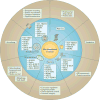
Figure 1. Mechanisms underlying deregulated ubiquitylation in cancer
Deregulated ubiquitylation in cancer can be attributed to epigenetic, genetic, transcriptional and post-translational mechanisms. Some ubiquitin ligases (E3s) are encoded by genes that are well recognized to confer susceptibility for familial cancers, such as the gene encoding von Hippel–Lindau disease tumour suppressor (VHL) in renal cell carcinoma (RCC) or the gene encoding BRCA1 in breast cancer and ovarian cancer. Large-scale analyses of cancer genomes have identified additional E3s that are altered by recurrent mutations or copy number changes in diverse cancers. The abundance and activity of E3s can also be regulated by post-translational mechanisms such as phosphorylation, ubiquitylation or protein–protein interactions, as demonstrated, for example, with the E3s MDM2 and SIAH2 (REFS 96,98,195,196). The activity and abundance of deubiquitylating enzymes (DUBs) are also regulated genetically and epigenetically, as recently reviewed. In addition to deregulation of E3s and DUBs, the ubiquitin system is modulated by genetic alterations of the targeted substrates. For example, recognition of the ubiquitylation sites on MYC and the fusion protein transmembrane protease serine 2 (TMPRSS2)–ETS-related gene (ERG) is disrupted by mutations. Ubiquitylation is a dynamic and reversible process that responds to a variety of internal and external stresses, including DNA damage and hypoxic, oxidative and metabolic stresses, which are all encountered by cancer cells during malignant transformation, during metastatic dissemination and in response to therapy. Each individual alteration in the ubiquitin system can have a profound effect on the regulation of cancer-associated pathways by modulating the localization, activity, signalling complex formation and abundance of major regulatory hubs. AKR1C3, aldo-keto reductase family 1 member C3; APC/C, anaphase-promoting complex; also known as the cyclosome; BAP1, BRCA1-associated protein 1; BTRC, encoding β-TRCP; CYLD, cylindromatosis; E2, ubiquitin-conjugating enzyme; FBXO11, F-box only protein 11; FBXW7, F-box/WD repeat-containing protein 7; GSK3, glycogen synthase kinase 3; HECT, homologous to E6AP carboxy terminus; KEAP1, kelch-like ECH-associated protein 1; NRF2, nuclear factor erythroid 2-related factor 2; PARK2, encoding parkin; RBR, RING-between-RING; RING, really interesting new gene; SIAH, seven in absentia homolgue; SKP2, S-phase kinase-associated protein 2; SPOP, speckle-type POZ protein; STUB1, STIP1 homology and U box-containing protein 1 (also known as CHIP); TRIM7, tripartite motif 7; U-box, UFD2 homology; Ub, ubiquitin; USP, ubiquitin carboxyl-terminal hydrolase.
Figure 2
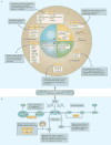
Figure 2. Cellular processes affected by deregulated ubiquitylation in cancer
Representative ubiquitin ligases (E3s) that are deregulated in cancer and the biological processes expected to be affected are depicted. As E3s ubiquitylate a diverse set of substrates, E3 loss or gain of function affects multiple cellular processes simultaneously. For example, S-phase kinase-associated protein 1 (SKP1)–cullin 1–F-box protein (SCF)–F-box/WD repeat-containing protein 7 (FBXW7) targets cell cycle regulators (for example, cyclin E), oncogenic transcription factors (for example, MYC), cell surface receptors (for example, NOTCH1), signalling molecules (for example, mTOR) and apoptosis regulators (for example, myeloid cell leukaemia 1 (MCL1)) for proteasomal degradation. Therefore, SCF–FBXW7 loss of activity through mutations or deletions leads to genomic instability, increased proliferation and survival and the rewiring of transcriptional and signalling programmes that affect cancer cell migration, metabolism and stemness. Similarly, SCF–β-transducin repeat-containing protein (β-TRCP) can serve as a signalling hub to coordinate increased protein synthesis and pro-survival signals following pro-growth stimuli in normal cells and in cancer cells. Thus, the β-TRCP signalling circuits provide a platform for therapeutic intervention, for example, in cancers characterized by activation of mTOR signalling. APC/C, anaphase-promoting complex; also known as the cyclosome; CRL3, cullin 3–really interesting new gene (RING)–E3 ligase; ER, endoplasmic reticulum; HRD1, also known as synoviolin; GP78, also known as AMFR; KEAP1, kelch-like ECH-associated protein 1; SIAH, seven in absentia homologue; SKP2, S-phase kinase-associated protein 2; SPOP, speckle-type POZ protein; STUB1, STIP1 homology and U box-containing protein 1; TRIM28, tripartite motif 28.
Figure 3
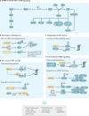
Figure 3. Ubiquitin ligases coordinate the cell cycle and DNA damage repair to maintain genome integrity
a | Phosphorylation and ubiquitylation coordinate the temporal activity of cyclin-dependent kinase (CDK)–cyclin complexes, thereby mediating cell cycle progression and checkpoint control. Displayed are some of the ubiquitin ligases (E3s), which are well-known cell cycle regulators, such as the APC/C (anaphase-promoting complex; also known as the cyclosome) and S-phase kinase-associated protein 1 (SKP1)–cullin 1–F-box protein (SCF)–F-box/WD repeat-containing protein 7 (FBXW7) complexes and parkin, whose function in the cell cycle is emerging. APC/C primarily mediates progression through mitosis by temporally coordinating the recruitment of co-activators (cell division cycle 20 (CDC20) or CDC20-like protein 1 (CDH1)). Thus, APC/C–CDC20 mediates anaphase entry, while APC/C–CDH1 acts during mitotic exit and early G1. APC/C is also regulated by phosphorylation, binding of inhibitory molecules such as early mitotic inhibitor 1 (EMI1; also known as FBXO5) and the spindle assembly checkpoint (SAC). SCF is a four-protein complex consisting of the scaffold cullin 1, a really interesting new gene (RING) domain-containing component RBX1, the SKP1 adaptor protein and one of ~68 various F-box proteins (for example, FBXW7, SKP2 or β-transducin repeat-containing protein (β-TRCP)). Most F-box proteins mediate the recognition of substrates by binding to their phosphodegron motifs (although phosphorylation-independent mechanisms of substrate recognition exist), which contain residues that can be phosphorylated by multiple kinases. The SCF complex is active throughout the cell cycle; SCF substrates include a subset of cyclins and CDK inhibitors, through which the SCF complex regulates progression from G1 to the onset of mitosis. Some example substrates of each E3 are displayed in the cream boxes, and the effects of altered E3 function in cancer are indicated in the grey boxes. b | DNA double strand breaks (DSBs) mediate activation of DNA damage sensors ataxia telangiectasia mutated (ATM), ataxia telangiectasia and Rad3-related (ATR), checkpoint kinase 1 (CHK1) and CHK2. These in turn inactivate the E3 MDM2, leading to p53 stabilization and/or promoting SCF–β-TRCP-mediated degradation of CDK phosphatases CDC25A, CDC25B and CDC25C; both pathways converge on the attenuation of CDK activity. ATM and ATR also initiate the recruitment of the DNA repair machinery to the sites of DNA damage, which is in turn under the control of ubiquitylation. Suppression of homologous recombination (HR) during G1 involves competition between p53-binding protein 1 (53BP1) and BRCA1 at DSBs, APC/C–CDH1-mediated CtBP-interacting protein (CTIP) degradation and cullin 3–RING–E3 ligase (CRL3)–kelch-like ECH-associated protein 1 (KEAP1)-dependent inhibition of BRCA1–partner and localizer of BRCA2 (PALB2)–BRCA2 complex formation. CEP68, centrosomal protein of 68 kDa; eEF2K, eukaryotic elongation factor 2 kinase; FANCM, Fanconi anaemia group M protein; NHEJ, non-homologous end joining; PLK1, polo-like kinase 1; REST, repressor-element 1 (REI)-silencing transcription factor; Ub, ubiquitin; USP11, ubiquitin carboxyl-terminal hydrolase 11.
Figure 4
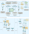
Figure 4. Regulation of mitotic signalling by ubiquitin ligases
a | Simplified schematics of the PI3K–AKT–mTOR and MAPK pathways are depicted. Ubiquitin ligases (E3s) and deubiquitylating enzymes (DUBs) regulate degradation, activity or complex assembly of receptor tyrosine kinases (RTKs), kinases and signalling molecules. b | Monoubiquitylation or diubiquitylation modulates RAS activity. Elevated expression of the DUB OTU domain-containing ubiquitin aldehyde-binding protein 1 (OTUB1) may contribute to sustained RAS activation, as demonstrated in lung cancer. c | High levels of the PI3K regulatory subunit p85β compete with active PI3K (made up of p85–p110 heterodimers) for substrate binding, thereby limiting PI3K activity. Following dephosphorylation of p85β by PTPL1, p85β is ubiquitylated by F-box and leucine-rich repeat protein 2 (FBXL2), leading to its proteasomal degradation and thereby increasing PI3K signalling output. d | PARK2 (which encodes E3 parkin) is frequently inactivated or downregulated in cancer by diverse mechanisms. Parkin loss indirectly increases AKT activity by affecting AKT upstream regulators. First, parkin loss leads to accumulation of parkin substrates, including the epidermal growth factor receptor (EGFR). Second, parkin loss impairs mitochondrial metabolism that results, from a cascade of events (indicated by the dotted line), in PTEN S-nitrosylation (by addition of the functional group S-nitrosothiol (SNO)), which primes PTEN for degradation. K63-linked polyubiquitylation of AKT by S-phase kinase-associated protein 1 (SKP1)–cullin 1–F-box protein (SCF)–SKP2 directly increases its activity. AKT can regulate stabilization and localization of SKP2, wherein AKT either directly phosphorylates SKP2 (REFS 89,90) (not shown) or mediates histone acetyltransferase p300 activation, which in turn leads to SKP2 acetylation (Ac) and thus stabilization and cytoplasmic retention of SKP2 (REF. 91). e | A casein kinase Iα (CKIα)–SCF–β-transducin repeat-containing protein (β-TRCP) auto-amplification loop mediates full activation of mTOR via DEP domain-containing mTOR-interacting protein (DEPTOR) degradation. The dynamic assembly of mTOR complex 1 (mTORC1) and mTORC2 is under the control of ubiquitylation. Ubiquitin-dependent deregulation of these signalling hubs in turn alters cellular processes as diverse as cell growth, metabolism, DNA repair, transcription, translation and survival. FOXO, forkhead box protein O; GSK3β, glycogen synthase kinase 3β; NF-κB, nuclear factor-κB; OTUD7B, OTU domain-containing protein 7B; P, phosphorylation; PIP, phosphatidylinositol phosphate; PRAS40, proline-rich AKT1 substrate 1; PROTOR, proline-rich protein; RABEX5, RAB5 GDP/GTP exchange factor; RAPTOR, regulatory-associated protein of mTOR; RICTOR, rapamycin-insensitive companion of mTOR; TRAF, tumour necrosis factor (TNF)-receptor-associated factor; TSC1, tuberous sclerosis 1 (also known as harmartin); TSC2, also known as tuberin; Ub, ubiquitin.
Figure 5
Figure 5. Ubiquitin ligases regulate the intrinsic and extrinsic apoptotic pathways
a | Cells maintain anti-apoptotic proteins at high levels, and pro-apoptotic BCL-2 homology domain 3 (BH3)-only proteins are transcriptionally upregulated mainly by stress-responsive transcription factors. This difference in protein abundance prevents oligomerization of the pro-apoptotic effector proteins BAX and BCL-2 antagonist/killer 1 (BAK1) and blocks mitochondrial outer membrane permeabilization (MOMP), thus inhibiting activation of caspase-dependent apoptosis. Increased mitogenic signalling can prevent apoptosis induction by targeting pro-apoptotic proteins for degradation, as exemplified by the S-phase kinase-associated protein 1 (SKP1)–cullin 1–F-box protein (SCF)–β-transducin repeat-containing protein (β-TRCP)-dependent degradation of pro-apoptotic BH3-only protein BIM (also known as BCL2L11) following ERK-mediated and ribosomal S6 kinase (RSK)-mediated phosphorylation of BIM. Consequently, knockdown of either β-TRCP or RSK induces apoptosis in non-small-cell lung cancer (NSCLC) cell lines. Similarly, downregulation of anti-apoptotic proteins by ubiquitin-mediated mechanisms, as illustrated here for myeloid cell leukaemia 1 (MCL1), can lower the threshold for apoptosis induction and sensitize cells to apoptosis-inducing insults. Decreased activity of MCL1-targeting ubiquitin ligases (E3s) in cancer therefore increases apoptosis resistance. b | Receptors of the tumour necrosis factor (TNF) superfamily are made up of TNF receptor 1 (TNFR1)-like (including TNFR1, DR3 (also known as TNFRSF25) and DR6 (also known as TNFRSF21)) and TNF-related apoptosis-inducing ligand (TRAIL) receptor (TRAILR)-like (including CD95 (also known as FAS and TNFRSF6) and TRAILR). Activation of TNFR1-like receptors recruits TNFR type 1-associated DEATH domain protein (TRADD), receptor-interacting serine/threonine-protein kinase 1 (RIPK1), TNF receptor-associated factor 2 (TRAF2), cellular inhibitor of apoptosis 1 (cIAP1) or cIAP2 and the linear ubiquitin chain assembly complex (LUBAC). K63-linked and M1-linked polyubiquitylation of RIPK1 by cIAPs and LUBAC, respectively, recruits inhibitor of nuclear factor-κB kinase (IKK), TAK1-binding protein (TAB) and TAK (also known as MAP3K7) to stimulate robust and rapid nuclear factor-κB (NF-κB) and MAPK signalling. TNFR1-induced cell death depends on receptor internalization and formation of a secondary, receptor-free cytoplasmic complex (complex II) composed of either RIPK1–FAS-associated death domain protein (FADD)–caspase 8 or RIPK1–RIPK3–mixed lineage kinase domain-like protein (MLKL), which mediates either apoptosis or necroptosis (a programmed form of necrosis), respectively. Formation of complex II is largely dependent on the activity of deubiquitylating enzymes (DUBs), specifically cylindromatosis (CYLD), A20 and ubiquitin thioesterase OTULIN. Alterations in the activity of these DUBs or of the E3s cIAP and LUBAC are reported in diverse cancers, where they lead to apoptosis resistance while stimulating oncogenic NF-κB signalling,. By contrast, stimulation of TRAILR-like primarily forms the death-inducing signalling complex containing FADD, caspase 8, caspase 10 and FLICE-like inhibitory protein (FLIP; also known as CFLAR). Complex II contains FADD, procaspase 8 filaments, RIPK1 and TRAF2. As indicated by the lighter shaded complex, RIPK1 and LUBAC may be directly recruited to complex I, allowing MAPK and NF-κB activation independent of complex II formation. Understanding the ubiquitin-mediated mechanisms that result in pro-tumorigenic NF-κB activation upon TRAILR stimulation is one of the important steps towards improving the therapeutic effect of TRAILR agonists, which are currently being evaluated as anticancer drugs. APC/C, anaphase-promoting complex; also known as the cyclosome; BCL-xL, also known as BCL2L1; BID, BH3, interacting domain death agonist; FBXW7, F-box/WD repeat-containing protein 7; HOIL1, haeme-oxidized IRP2 ubiquitin ligase 1 (also known as RBCK1); HOIP, HOIL1-interacting protein (also known as RNF31); NEMO, NF-κB essential modulator; P, phosphorylation; PUMA, p53 up-regulated modulator of apoptosis (also known as BBC3); SHARPIN, shank-associated RH domain-interacting protein; Ub, ubiquitin.

References

    1. Gross S, Rahal R, Stransky N, Lengauer C, Hoeflich KP. Targeting cancer with kinase inhibitors. J. Clin. Invest. 2015;125:1780–1789. - PMC - PubMed
    1. Duan G, Walther D. The roles of post-translational modifications in the context of protein interaction networks. PLoS Comput. Biol. 2015;11:e1004049. - PMC - PubMed
    1. Ciechanover A. The unravelling of the ubiquitin system. Nat. Rev. Mol. Cell Biol. 2015;16:322–324. This is a timely updated essay on the fundamentals of the ubiquitin system. - PubMed
    1. Foot N, Henshall T, Kumar S. Ubiquitination and the regulation of membrane proteins. Physiol. Rev. 2017;97:253–281. - PubMed
    1. Khaminets A, Behl C, Dikic I. Ubiquitin-dependent and independent signals in selective autophagy. Trends Cell Biol. 2016;26:6–16. This is a detailed review on the regulation of autophagy by ubiquitin-related and non-related pathways. - PubMed

Publication types