. 2012 Oct 29;2(31):11876-11886.
doi: 10.1039/C2RA21544F.

Highly lipophilic fluorescent dyes in nano-emulsions: towards bright non-leaking nano-droplets

Affiliations

Highly lipophilic fluorescent dyes in nano-emulsions: towards bright non-leaking nano-droplets

Andrey S Klymchenko et al. RSC Adv. .

Abstract

Dye-loaded lipid nano-droplets present an attractive alternative to inorganic nanoparticles, as they are composed of non-toxic biodegradable materials and easy to prepare. However, to achieve high fluorescence brightness, the nano-droplets have to be heavily loaded with the dyes avoiding fluorescence self-quenching and release (leakage) of the encapsulated dyes from the nano-droplets in biological media. In the present work, we have designed highly lipophilic fluorescent derivatives of 3-alkoxyflavone (F888) and Nile Red (NR668) that can be encapsulated in the lipophilic core of stable nano-emulsion droplets at exceptionally high concentrations in the oil core, i.e. up to 170 mM and 17 mM, respectively, corresponding to ~ 830 and 80 dyes per 40-nm droplet. Despite this high loading, these dyes keep high fluorescence quantum yield and thus, provide high nano-droplet brightness, probably due to their bulky structure preventing self-quenching. Moreover, simultaneous encapsulation of both dyes at high concentrations in single nano-droplets allows observation of FRET. FRET and fluorescence correlation spectroscopy (FCS) studies showed that NR668 release in the serum-containing medium is very slow, while the reference hydrophobic dye Nile Red leaks immediately. This drastic difference in the leakage profile between NR668 and Nile Red was confirmed by in vitro cellular studies as well as by in vivo angiography imaging on zebrafish model, where the NR668-loaded nano-droplets remained in the blood circulation, while the parent Nile Red leaked rapidly from the droplets distributing all over the animal body. This study suggests new molecular design strategies for obtaining bright nano-droplets without dye leakage and their use as efficient and stable optical contrast agents in vitro and in vivo.

Keywords: Förster resonance energy transfer; dye release; fluorescence correlation spectroscopy; fluorescent lipid nano-droplets; intra-vital imaging; nano-emulsions.


Figures

Figure 1
Figure 1
Schematic presentation of a nano-droplet and chemical structure of the new lipophilic dyes used for encapsulation. Nile Red was used as a reference for characterizing the release properties.
Figure 2
Figure 2
Fluorescence spectra of F888 and NR668 in nano-emulsions, neat Labrafac CC® oil and ethanol.
Figure 3
Figure 3
Typical examples of FCS correlation curves (A) and obtained nano-droplet brightness (B) for dye-loaded nano-emulsions compared to TMR, used as reference. Inset: principle of FCS measurements.
Figure 4
Figure 4
Observation of FRET inside lipid nano-droplets at different concentrations of donor (F888) and acceptor (NR668), encapsulated at 1/1 molar ratio. Dotted line corresponds to nano-droplets containing only the donor. Fluorescence spectra were recorded at 390 nm excitation wavelength. To compare the spectra, their absolute fluorescence intensity was divided by the F888 concentration.
Figure 5
Figure 5
Investigation by FRET of the dye release from lipid nano-droplets in different media: water, Opti-MEM (OM) and Opti-MEM with 10 vol.% FBS (OM+FBS). Droplets encapsulating 0.5% of F888 (with respect to Labrafac CC®) as energy donor and 0.5 wt.% of Nile Red (A) or NR668 (B) as energy acceptor were used. The nano-droplets were diluted 10000-times from the original formulation into the medium of interest. The first fluorescence spectra were measured after 3 min incubation at RT (A and B). Then, samples were incubated for 1 h, 3 h and 6 h at 37°C. FRET was quantified as the fluorescence intensity ratio between the maximum of the donor (450 nm) and acceptor (590 nm) (C). The donor in the nano-droplets was excited at 390 nm.
Figure 6
Figure 6
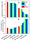
Dye release studies by FCS. Brightness and apparent concentration of emissive species in water and in biological media determined from FCS data. Brightness is the photon count rate per droplet. The nanoemulsions prepared in water were measured 5 min after dilution in the indicated media at room temperature or after incubation in these media at 37 °C for 1h or 6h. Data in water, measured 5 min after dilution at room temperature are presented for comparison.
Figure 7
Figure 7
Combined fluorescence and transmission images of HeLa cells incubated with nano-droplets containing 0.1 wt.% of Nile Red (A and B) or 1 wt.% of NR668 (C and D) for different times: 15 min (A and C) and 2 h (B and D).
Figure 8
Figure 8
Zebrafish microangiography using nano-droplets containing 0.1 wt.% of Nile Red (A, B and C) or 1 wt.% of NR668 (D, E and F) with the Tg(fli1:eGFP)y1 line. 3 days post fertilization, the living zebrafish embryos were injected 2.3 nL of nano-emulsions diluted twice in HEPES buffer (pH 7.4) and imaged with a confocal microscope. Images present the global view (upper panels) and a zoom in of the trunk vasculature (lower panels). The images in green (A and D) present endothelial cells expressing eGFP, while in red – fluorescence of Nile Red (B) and NR668 (E), 30 min after the injection of the nano-emulsion. The arrows show the endothelial cells. The merged image (C) shows the localization of Nile Red in the endothelium and the global diffuse labeling of the entire embryo, while the merged image (F) shows no colocalization of NR688 with the endothelial cells.

References

    1. Brigger I, Dubernet C, Couvreur P. Adv Drug Deliv Rev. 2002;54:631. - PubMed
    1. Horn D, Rieger J. Angew Chem Int Ed. 2001;40:4330. - PubMed
    1. Liong M, Lu J, Kovochich M, Xia T, Ruehm SG, Nel AE, Tamanoi F, Zink JI. ACS Nano. 2008;2:889. - PMC - PubMed
    1. Sharma P, Brown S, Walter G, Santra S, Moudgil B. Adv Colloid Interface Sci. 2006;123-126:471. - PubMed
    1. Lu J, Liong M, Zink JI, Tamanoi F. Small. 2007;3:1341. - PubMed

LinkOut - more resources