

As a Definitive Choice of Treatment, Joint and Defect Spanning Multiplanar Tubular External Fixation in the Management of Pediatric Open Defective Supracondylar Humerus Fracture: A Case Report
- PMID: 29242803
- PMCID: PMC5728008
- DOI: 10.13107/jocr.2250-0685.908
As a Definitive Choice of Treatment, Joint and Defect Spanning Multiplanar Tubular External Fixation in the Management of Pediatric Open Defective Supracondylar Humerus Fracture: A Case Report
Abstract
Introduction: Although supracondylar fractures of the humerus are common in children, open fractures of are extremely rare injuries. Gustilo- Anderson Type-III fractures in the upper extremity are primarily associated with considerable bone loss resulting from high energy trauma. In this study, a case of open pediatric supracondylar humerus fracture treated by a joint and defect spanning multiplanar tubular external fixation is presented.
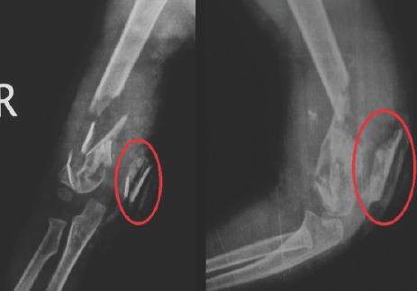
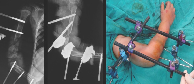
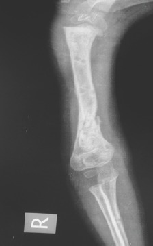
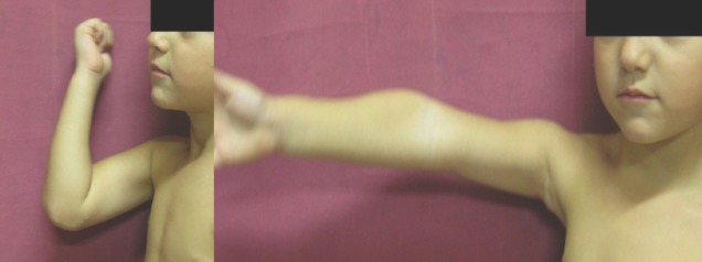
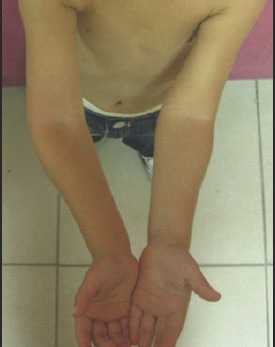
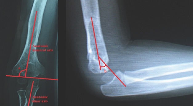
Case report: A boy aged 3 years suffered from a Gustilo-Anderson Type-IIIA supracondylar humerus fractures. There was no neurovascular compromise. Intravenous antibiotic regimen was introduced. A joint and defect spanning multiplanar tubular external fixation was employed within 4 h of the accident. After the operation, the patient was closely followed in the inpatients unit. The external fixator was removed in the 6th week of follow-up, and range of motion exercises was started. With the exception of limited flexion of the elbow in 12th month of follow-up, supination and pronation were full. At the 48th month follow-up, 120° of elbow flexion, full extension, and full forearm rotation were observed. Physical examination showed 15° change in carrying angle to cubitus varus; radiologic examination showed a slight varus angulation of the distal humerus, a decreased Baumann's angle. The Mayo elbow performance score was 100 points.
Conclusion: When taking into account the high remodeling capacity, healing potential, and greater resistance to joint stiffness in fractures of children, joint and external fixators appear as a viable definitive treatment in such cases.
Keywords: Open pediatric fracture; bone loss; external fixator; supracondylar humerus.
Conflict of interest statement
Conflict of Interest: Nil
Figures
Similar articles
-
Can a linear external fixator stand as a surgical alternative to open reduction in treating a high-grade supracondylar humerus fracture?J Int Med Res. 2019 Jan;47(1):133-141. doi: 10.1177/0300060518797022. Epub 2018 Sep 10. J Int Med Res. 2019. PMID: 30198367 Free PMC article.
-
[Case control study on micro external fixator in treating supracondylar fracture of humerus in children].Zhongguo Gu Shang. 2020 Oct 25;33(10):902-6. doi: 10.12200/j.issn.1003-0034.2020.10.003. Zhongguo Gu Shang. 2020. PMID: 33107250 Chinese.
-
Results of displaced supracondylar humerus fractures treated with open reduction and internal fixation after a mean 22.4 years of follow-up.J Shoulder Elbow Surg. 2015 Apr;24(4):640-6. doi: 10.1016/j.jse.2014.12.010. Epub 2015 Jan 31. J Shoulder Elbow Surg. 2015. PMID: 25648970
-
Operative treatment of supracondylar fractures of the humerus in children: the Cincinnati experience.Acta Orthop Belg. 1996;62 Suppl 1:41-50. Acta Orthop Belg. 1996. PMID: 9084559 Review.
-
Open fractures of the upper limb - do the BOAST guidelines need an update?Injury. 2023 Apr 15:S0020-1383(23)00374-1. doi: 10.1016/j.injury.2023.04.029. Online ahead of print. Injury. 2023. PMID: 37080881 Review.
Cited by
-
Management of Open Supracondylar Humeral Fracture in Children: A Case Report and Literature Review.Cureus. 2023 Nov 1;15(11):e48119. doi: 10.7759/cureus.48119. eCollection 2023 Nov. Cureus. 2023. PMID: 38046701 Free PMC article.
References
-
- Haasbeek JF, Cole WG. Open fractures of the arm in children. Bone JointJ. 1995;77:576–81. - PubMed
-
- Lewine E, Kim JM, Miller PE, Waters PM, Mahan ST, Snyder B, et al. Closed versus open supracondylar fractures of the humerus in children: A Comparison of clinical and radiographic presentation and results. J Pediatr Orthop. 2016 - PubMed
-
- Wiley JJ. Current Concepts of Bone Fragility. Berlin: Springer-Verlag; 1986. Fracture patterns in children; p. 159.
-
- Stewart DG, Jr, Kay RM, Skaggs DL. Open fractures in children. Principles of evaluation and management. J Bone Joint Surg Am. 2005;87:2784–98. - PubMed
-
- Luhmann SJ, Schootman M, Schoenecker PL, Dobbs MB, Gordon JE. Complications and outcomes of open pediatric forearm fractures. J Pediatr Orthop. 2004;24:1–6. - PubMed
Publication types
LinkOut - more resources
Full Text Sources
Research Materials