Review
. 2018 Jan;472(1):159-172.
doi: 10.1007/s00428-017-2277-x. Epub 2017 Dec 14.

Gut microbiome: a new player in gastrointestinal disease

Affiliations
Review

Gut microbiome: a new player in gastrointestinal disease

Gregor Gorkiewicz et al. Virchows Arch. 2018 Jan.

Abstract

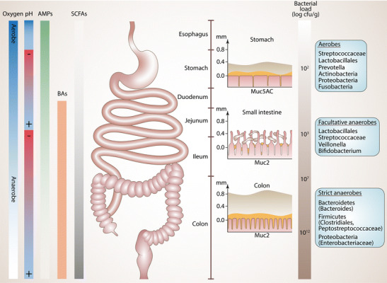
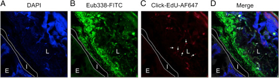
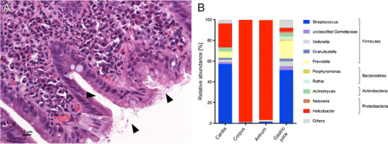
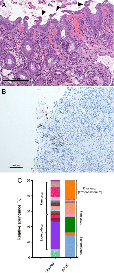
The gastrointestinal (GI) tract harbors a diverse and host-specific gut microbial community. Whereas host-microbe interactions are based on homeostasis and mutualism, the microbiome also contributes to disease development. In this review, we summarize recent findings connecting the GI microbiome with GI disease. Starting with a description of biochemical factors shaping microbial compositions in each gut segment along the longitudinal axis, improved histological techniques enabling high resolution visualization of the spatial microbiome structure are highlighted. Subsequently, inflammatory and neoplastic diseases of the esophagus, stomach, and small and large intestines are discussed and the respective changes in microbiome compositions summarized. Finally, approaches aiming to restore disturbed microbiome compositions thereby promoting health are discussed.

Keywords: Carcinogenesis; Dysbiosis; Fecal microbiome transplantation; Gut microbiome; Inflammation; Spatial microbiome organization.


Conflict of interest statement

The authors declare that they have no conflicts of interest.

Figures

Fig. 1
Fig. 1
Biogeography and factors shaping the spatial organization of the gut microbiome. Left: factors determining gut segment specific microbiome composition like oxygen, pH, bile acids (BA), antimicrobial peptides (AMPs), and concentration of short-chain fatty acids (SCFAs). Middle: schematic representation of the GI tract and of the segment specific mucus layer architecture (adapted from [33, 34]). The inner solid (amber) and the outer loose mucus layer (gray) are shown. Note that in the stomach and colon the mucus layer is continuous, whereas in the small intestine the layer is discontinuous. Muc5AC and Muc2 denote the dominant mucins produced in the respective gut segment. Right: bacterial load and typical taxonomic compositions of different gut segments
Fig. 2
Fig. 2
In vivo tracking and improved spatial resolution of gut-microbiome interactions. A mouse colon was fixed in Carnoy’s solution to preserve mucus layer architecture. a The section was counterstained with 4′,6-diamidino-2-phenylindole (DAPI) indicating the colonic epithelium (E) in the left lower corner, the interlaced (I) mucus layer (indicated by the two dotted lines), which is devoid of bacteria, and the colonic lumen (L) on the right side. The structure in the upper right depicts a plant component. b Bacteria were stained by FISH using a fluorescein isothiocyanate (FITC) pan-specific EUB338 probe (green) which covers approximately 90% of the domain bacteria. c In this experiment, mice were gavaged with the gut bacterium Alistipes finegoldii (phylum Bacteroidetes), which were cultured in the presence of the thymidine analog 5-ethynyl-2′-deoxyuridine (EdU). Metabolically active Alistipes was tracked utilizing click chemistry that is based on a copper-catalyzed covalent reaction between an alkyne (within the EdU) and an AlexaFluor® 647-containing azide. Note that the bacterium colonizes the luminal site of the colon, not the mucus layer (arrows). d The right picture shows the merged panels. Cells were imaged on a Zeiss Axioobserver Z1 microscope equipped with a LSM700 confocal unit. Original magnification 400×
Fig. 3
Fig. 3
Stomach microbiome in chronic H. pylori gastritis. a Gastric corpus biopsy signifying the preferred mucosal niche of H. pylori (arrow heads). b Microbiome analysis (based on the 16S rRNA gene) indicates the dominance of H. pylori at the mucosal sites, whereas in gastric juice only few H. pylori are prevalent
Fig. 4
Fig. 4
Histology and microbiome representation of antibiotic-associated hemorrhagic colitis (AAHC). a Colon histology with micropapillary epithelial protrusions (arrow heads) indicating the cytotoxic effect of the enterotoxin tilivalline produced by K. oxytoca. b Activated caspase-3 immunohistochemistry signifying epithelial cell apoptosis. c Fecal microbiome composition in AAHC (based on the 16S rRNA gene analysis). A highly reduced overall diversity is evident with the overgrowth of the proteobacterium K. oxytoca. A diverse healthy fecal microbiome is shown on the left

References

    1. Lynch SV, Pedersen O. The human intestinal microbiome in health and disease. N Engl J Med. 2016;375(24):2369–2379. - PubMed
    1. Bordenstein SR, Theis KR. Host biology in light of the microbiome: ten principles of holobionts and hologenomes. PLoS Biol. 2015;13(8):e1002226. - PMC - PubMed
    1. The Human Microbiome Project Consortium Structure, function and diversity of the healthy human microbiome. Nature. 2012;486(7402):207–214. - PMC - PubMed
    1. Qin J, Li R, Raes J, Arumugam M, Burgdorf KS, Manichanh C, Nielsen T, Pons N, Levenez F, Yamada T, Mende DR, Li J, Xu J, Li S, Li D, Cao J, Wang B, Liang H, Zheng H, Xie Y, Tap J, Lepage P, Bertalan M, Batto JM, Hansen T, Le Paslier D, Linneberg A, Nielsen HB, Pelletier E, Renault P, Sicheritz-Ponten T, Turner K, Zhu H, Yu C, Li S, Jian M, Zhou Y, Li Y, Zhang X, Li S, Qin N, Yang H, Wang J, Brunak S, Doré J, Guarner F, Kristiansen K, Pedersen O, Parkhill J, Weissenbach J, MetaHIT Consortium. Bork P, Ehrlich SD, Wang J. A human gut microbial gene catalogue established by metagenomic sequencing. Nature. 2010;464(7285):59–65. - PMC - PubMed
    1. Halwachs B, Madhusudhan N, Krause R, Nilsson RH, Moissl-Eichinger C, Högenauer C, Thallinger GG, Gorkiewicz G. Critical issues in mycobiota analysis. Front Microbiol. 2017;8:180. - PMC - PubMed

MeSH terms

Grants and funding

LinkOut - more resources