. 2017 Dec 15:6:e31661.
doi: 10.7554/eLife.31661.

Diverse functions of homologous actin isoforms are defined by their nucleotide, rather than their amino acid sequence

Affiliations

Diverse functions of homologous actin isoforms are defined by their nucleotide, rather than their amino acid sequence

Pavan Vedula et al. Elife. .

Abstract

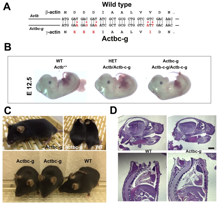
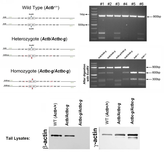
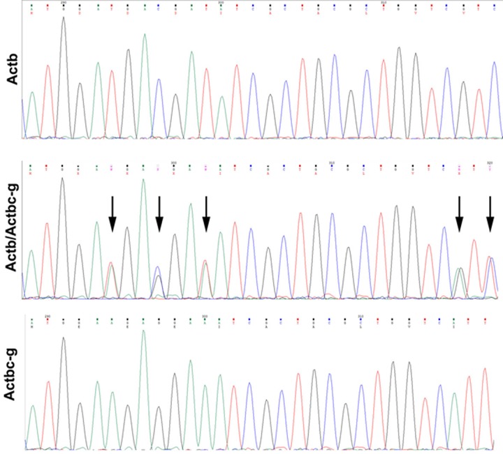
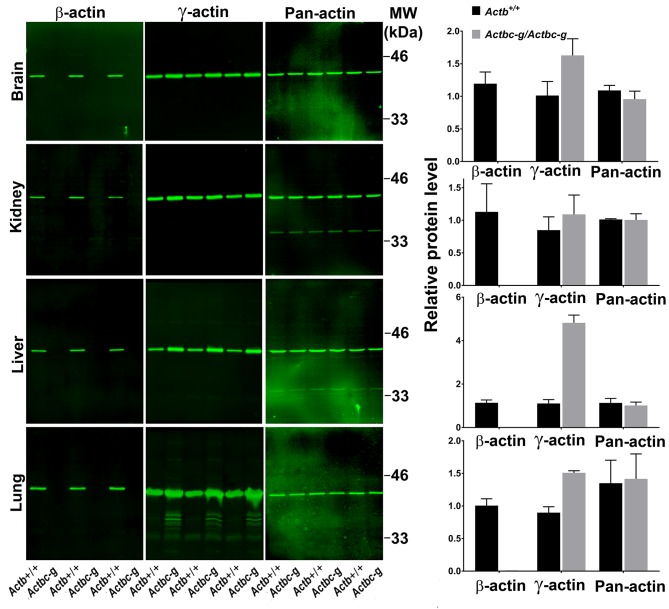
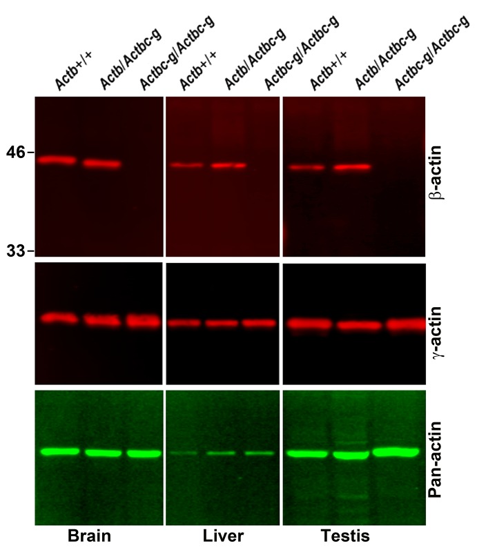
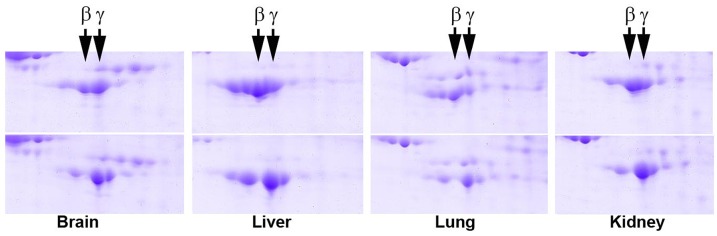
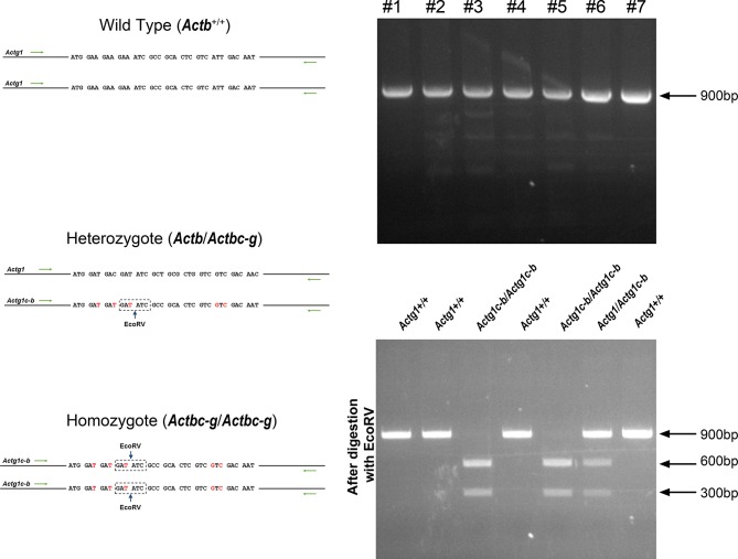
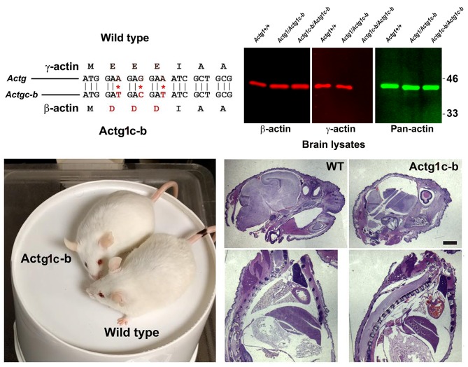
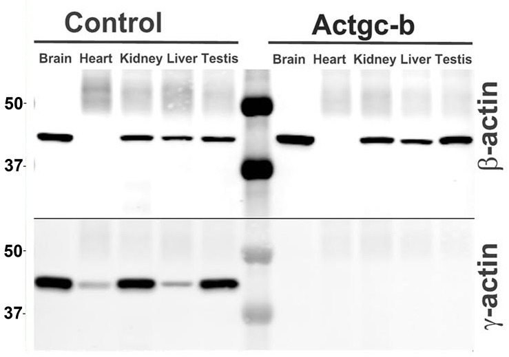
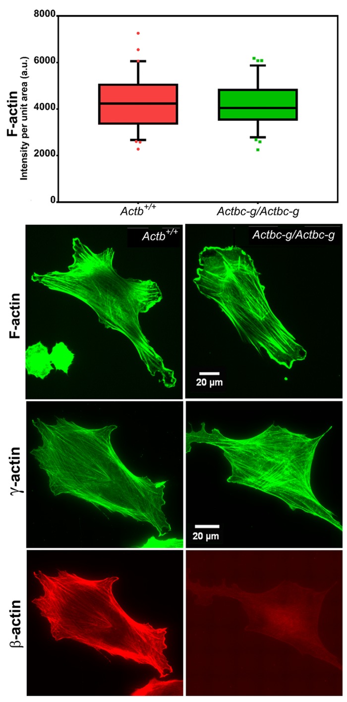
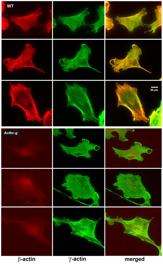
β- and γ-cytoplasmic actin are nearly indistinguishable in their amino acid sequence, but are encoded by different genes that play non-redundant biological roles. The key determinants that drive their functional distinction are unknown. Here, we tested the hypothesis that β- and γ-actin functions are defined by their nucleotide, rather than their amino acid sequence, using targeted editing of the mouse genome. Although previous studies have shown that disruption of β-actin gene critically impacts cell migration and mouse embryogenesis, we demonstrate here that generation of a mouse lacking β-actin protein by editing β-actin gene to encode γ-actin protein, and vice versa, does not affect cell migration and/or organism survival. Our data suggest that the essential in vivo function of β-actin is provided by the gene sequence independent of the encoded protein isoform. We propose that this regulation constitutes a global 'silent code' mechanism that controls the functional diversity of protein isoforms.

Keywords: actin; biochemistry; cell biology; coding sequence; isoforms; mouse.


Conflict of interest statement

PV, SK, NL, YW, SS, JW, SS, DD, AK No competing interests declared

Figures

Figure 1.
Figure 1.. β -coded γ -actin (Actbc-g) mice exhibit no phenotypic changes compared to control.
(A) CRISPR/Cas9 editing strategy used to generate Actbc-g mouse. (B) photos of Actbc-g E12.5 mouse embryos, with genotypes indicated. (C) photos of Actbc-g mice after gene editing, alone (top left) and next to age-matched (top right) and littermate wild type (WT) (bottom). Three mice from two different litters are shown. (D) H&E-stained sagittal sections of the heads (top) and bodies (bottom) of littermate P0 wild type (WT) and Actbc-g mice. Scale bar, 1 mm.
Figure 1—figure supplement 1.
Figure 1—figure supplement 1.. Generation of Actbc-g mouse.
Top left, genotyping strategy: editing of the N-terminal codons of the β-actin gene abolishes an EcoRV restriction site, enabling the screening of the edited gene variants by EcoRV digestion of the PCR-generated DNA fragments corresponding to the 5′ of the actin sequence. Top right, PCR products before (top) and after (bottom) EcoRV digestion. Bottom, western blots of wild type, heterozygous, and Actbc-g mouse tail lysates with antibodies to β- and γ- actin.
Figure 1—figure supplement 2.
Figure 1—figure supplement 2.. Sequencing result for wild type Actb and the edited Actbc-g alleles.
Screen shots from the Chromas sequence viewing software.
Figure 2.
Figure 2.. Actb gene editing abolishes β-actin protein from multiple organs and is accompanied by up-regulation of γ-actin without changing the total actin levels.
Western blot analysis showing images (left) and quantifications (right) of whole tissue lysates from wild type (Actb+/+) and Actbcg mice. Fluorescence images obtained from the Odyssey gel imager are shown. For quantification, total fluorescence from the 43 kDa actin band was normalized to the loading control and to the actin level in the first lane for each blot. Error bars represent SEM, n = 3.
Figure 2—figure supplement 1.
Figure 2—figure supplement 1.. Actin levels in Actbc-g mice are similar to control.
Western blots of wild type, heterozygous, and Actbc-g mouse brain lysates probed with antibodies to β- and γ-actin and total actin (pan-actin). Mouse genotypes are indicated on top of each lane, and the antibodies used are listed below each blot.
Figure 2—figure supplement 2.
Figure 2—figure supplement 2.. 2D gel distribution of actin isoforms in wild type (top) and Actbc-g (bottom) mouse tissue lysates.
Figure 2—figure supplement 3.
Figure 2—figure supplement 3.. Generation of Actg1c-b mouse.
Top, genotyping strategy: editing of the N-terminal codons of theγ- actin gene generates an EcoRV restriction site, enabling the screening of the edited gene variants by EcoRV digestion of the PCR-generated DNA fragments corresponding to the beginning of the actin sequence. Bottom, PCR products before (top) and after (bottom) EcoRV digestion.
Figure 2—figure supplement 4.
Figure 2—figure supplement 4.. γ-coded β-actin (Actg1c-b mice exhibit no phenotypic changes compared to control.
Top, CRISPR/Cas9 editing strategy used to generate Actg1c-b mouse. Bottom left, photos of Actg1c-b mouse after gene editing, to age-matched wild type. Bottom right, H and E-stained sagittal sections of the heads (top) and bodies (bottom) of littermate wild type (WT) and Actg1c-b mice. Scale bar, 1 mm.
Figure 2—figure supplement 5.
Figure 2—figure supplement 5.. Partial editing of the γ-actin gene to encode β-actin-like protein abolishes γ-actin protein from multiple organs.
Western blots with the actin antibodies indicated on the left using tissue homogenates from wild type control and Actg1c-b mouse.
Figure 3.
Figure 3.. Mouse embryonic fibroblasts derived from Actbc-g mice have normal actin cytoskeleton, despite complete lack of β-actin.
Top, quantification of total F-actin detected by Phalloidin-AlexaFluor488 staining of wild type (Actb+/+) and Actbc-g primary mouse embryonic fibroblasts. Numbers were averaged from 69 cells in WT and 76 cells in Actbc-g, obtained from two different primary cultures independently derived from two different littermate embryos for each set. Bottom, representative images of both cell types stained with Phalloidin-AlexaFluor488 or antibodies to both actin isoforms as indicated.
Figure 4.
Figure 4.. Mouse embryonic fibroblasts show no major changes in morphology and actin distribution.
Representative images of wild type (WT) and Actbc-g primary mouse embryonic fibroblasts stained with antibodies to both actin isoforms as indicated.
Figure 5.
Figure 5.. Mouse embryonic fibroblasts derived from Actbc-g mice migrate at normal rates.
Left, phase contrast images of the first (0′) and last (600′) frame taken from a representative time lapse videos of the WT and Actbc-g cells at the edge of a monolayer migrating into an infinite scratch wound. Overlay of the two frames is shown in the bottom row. Scale bar, 100 µm. Right top, quantification of the cell migration rate as μm2/min, WT: n = 28, Actbc-g: n = 29 averaged from two independently derived primary cultures for each set. See Supplemental Videos 1 and 2. Right bottom, quantification of cell directionality in single cell migration assays (calculated as persistence over time, WT: n = 49, Actbc-g: n = 50) suggests that single cell migration was not affected in Actbc-g cells.

Comment in

  • Fundamental differences.
    Gunning PW, Hardeman EC. Gunning PW, et al. Elife. 2018 Feb 1;7:e34477. doi: 10.7554/eLife.34477. Elife. 2018. PMID: 29388551 Free PMC article.

References

    1. Belyantseva IA, Perrin BJ, Sonnemann KJ, Zhu M, Stepanyan R, McGee J, Frolenkov GI, Walsh EJ, Friderici KH, Friedman TB, Ervasti JM. Gamma-actin is required for cytoskeletal maintenance but not development. PNAS. 2009;106:9703–9708. doi: 10.1073/pnas.0900221106. - DOI - PMC - PubMed
    1. Bergeron SE, Zhu M, Thiem SM, Friderici KH, Rubenstein PA. Ion-dependent polymerization differences between mammalian beta- and gamma-nonmuscle actin isoforms. Journal of Biological Chemistry. 2010;285:16087–16095. doi: 10.1074/jbc.M110.110130. - DOI - PMC - PubMed
    1. Bunnell TM, Burbach BJ, Shimizu Y, Ervasti JM. β-Actin specifically controls cell growth, migration, and the G-actin pool. Molecular Biology of the Cell. 2011;22:4047–4058. doi: 10.1091/mbc.E11-06-0582. - DOI - PMC - PubMed
    1. Bunnell TM, Ervasti JM. Delayed embryonic development and impaired cell growth and survival in Actg1 null mice. Cytoskeleton. 2010;67:564–572. doi: 10.1002/cm.20467. - DOI - PMC - PubMed
    1. Burgess-Cassler A, Johansen JJ, Santek DA, Ide JR, Kendrick NC. Computerized quantitative analysis of coomassie-blue-stained serum proteins separated by two-dimensional electrophoresis. Clinical Chemistry. 1989;35:2297–2304. - PubMed

Publication types