Review
. 2017 Dec 15;10(12):1429.
doi: 10.3390/ma10121429.

Potential of Bioactive Glasses for Cardiac and Pulmonary Tissue Engineering

Affiliations
Review

Potential of Bioactive Glasses for Cardiac and Pulmonary Tissue Engineering

Saeid Kargozar et al. Materials (Basel). .

Abstract

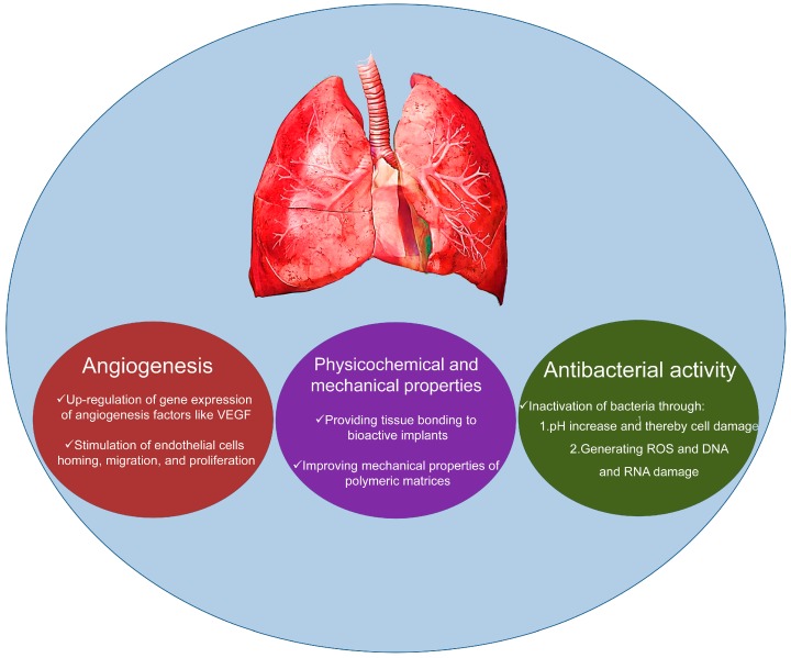
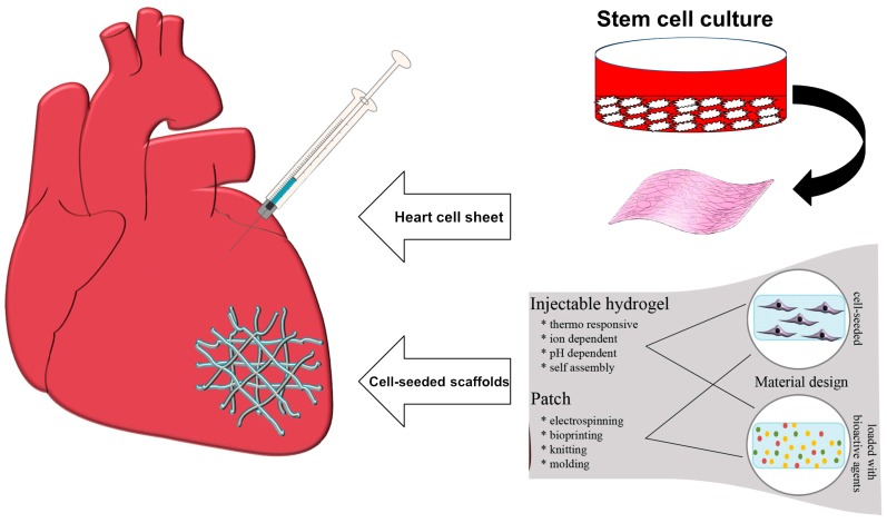
Repair and regeneration of disorders affecting cardiac and pulmonary tissues through tissue-engineering-based approaches is currently of particular interest. On this matter, different families of bioactive glasses (BGs) have recently been given much consideration with respect to treating refractory diseases of these tissues, such as myocardial infarction. The inherent properties of BGs, including their ability to bond to hard and soft tissues, to stimulate angiogenesis, and to elicit antimicrobial effects, along with their excellent biocompatibility, support these newly proposed strategies. Moreover, BGs can also act as a bioactive reinforcing phase to finely tune the mechanical properties of polymer-based constructs used to repair the damaged cardiac and pulmonary tissues. In the present study, we evaluated the potential of different forms of BGs, alone or in combination with other materials (e.g., polymers), in regards to repair and regenerate injured tissues of cardiac and pulmonary systems.

Keywords: angiogenesis; bioactive glasses; cardiac regeneration; lung tissue engineering; scaffold; soft tissue engineering.


Conflict of interest statement

The authors declare no conflict of interest.

Figures

Figure 1
Figure 1
Schematic representation of potential of BGs for cardiac and pulmonary tissue engineering.
Figure 2
Figure 2
Examples of two approaches to cardiac tissue engineering. Stem cells are differentiated into cardiac repair cells and seeded onto a scaffold material or mixed with an injectable matrix. The scaffold with the repair cells is placed at the site of MI or used to replace the infarcted tissue. Cells encapsulated within a hydrogel matrix are delivered to the site of injury via direct injection. Images from Hinderer et al. [66] © Wiley Publishers.
Figure 3
Figure 3
SEM micrographs of (left) gelatin/collagen scaffolds and (right) gelatin/collagen/BG scaffolds for cardiac tissue repair. Top: Pictures of bare scaffolds; middle: Cells seeded on the scaffolds; bottom: Nuclei staining of cells grown on scaffolds 15 days post cell seeding. Images from Barabadi et al. [74] © Elsevier.
Figure 4
Figure 4
Cross-sectional SEM images of elastomeric biomaterials (silicones containing or not 10 wt % bioactive glass (BG45S5) particles) for cardiac tissue repair: (a) pure silicone; (b) silicone with nano-sized bioactive glass (nano-BG); (c) silicone with BG microparticles by Schott (Schott-BG); and (d) silicone with BG microparticles by Mo-Sci-Corporation (Mo-Sci-BG); and (eg) the respective BG-containing composite films after four weeks of immersion in simulated body fluid (SBF). Images from Cohrs et al. [75] © Springer.
Figure 5
Figure 5
PDLLA/45S5 Bioglass® scaffolds for possible application in lung tissue engineering: SEM micrographs showing the microstructure of the polymeric foam filled with 40 wt % of glass in sections: (a) orthogonal to pore direction; and (b) parallel to pore direction; and A549 cells seeded on the same scaffolds and incubated for: (c) three days; and (d) six days. Images from Verrier et al. [85] © Elsevier.
Figure 6
Figure 6
Tubular PLGA/45S5 Bioglass® composite constructs: SEM micrographs showing: a radial section of an uncoated PLGA tube (a); and a detail of the cross-section (b); and the radial section of the composite tubular foam (c); and detail of the Bioglass® particles on a fracture surface (d). Images from Boccaccini et al. [86] © Elsevier.

References

    1. Dawber T.R., Moore F.E., Mann G.V., II Coronary Heart Disease in the Framingham Study. Int. J. Epidemiol. 2015;44:1767–1780. doi: 10.1093/ije/dyv346. - DOI - PubMed
    1. Galiè N., Humbert M., Vachiery J.-L., Gibbs S., Lang I., Torbicki A., Simonneau G., Peacock A., Vonk Noordegraaf A., Beghetti M., et al. 2015 ESC/ERS guidelines for the diagnosis and treatment of pulmonary hypertension: The joint task force for the diagnosis and treatment of pulmonary hypertension of the European Society of Cardiology (ESC) and the European Respiratory Society (ERS): Endorsed by: Association for European Paediatric and Congenital Cardiology (AEPC), International Society for Heart and Lung Transplantation (ISHLT) Eur. Heart J. 2015;37:67–119. - PubMed
    1. Members W.G., Go A.S., Mozaffarian D., Roger V.L., Benjamin E.J., Berry J.D., Blaha M.J., Dai S., Ford E.S., Fox C.S., et al. Heart disease and stroke statistics—2014 update: A report from the American Heart Association. Circulation. 2014;129:e28–e292. - PMC - PubMed
    1. Hashimoto K., Miyoshi K., Mizutani H., Otani S., Sugimoto S., Yamane M., Oto T. Successful Lung Transplantation for Pulmonary Disease Associated With Erdheim-Chester Disease. Ann. Thorac. Surg. 2017;104:e13–e15. doi: 10.1016/j.athoracsur.2017.02.020. - DOI - PubMed
    1. Levine D.J., Glanville A.R., Aboyoun C., Belperio J., Benden C., Berry G.J., Hachem R., Hayes D., Neil D., Reinsmoen N.L., et al. Antibody-mediated rejection of the lung: A consensus report of the International Society for Heart and Lung Transplantation. J. Heart Lung Transplant. 2016;35:397–406. doi: 10.1016/j.healun.2016.01.1223. - DOI - PubMed

LinkOut - more resources