Identification and characterization of the three members of the CLC family of anion transport proteins in Trypanosoma brucei
- PMID: 29244877
- PMCID: PMC5731698
- DOI: 10.1371/journal.pone.0188219
Identification and characterization of the three members of the CLC family of anion transport proteins in Trypanosoma brucei
Abstract
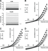
CLC type anion transport proteins are homo-dimeric or hetero-dimeric with an integrated transport function in each subunit. We have identified and partially characterized three members of this family named TbVCL1, TbVCL2 and TbVCL3 in Trypanosoma brucei. Among the human CLC family members, the T. brucei proteins display highest similarity to CLC-6 and CLC-7. TbVCL1, but not TbVCL2 and TbVCL3 is able to complement growth of a CLC-deficient Saccharomyces cerevisiae mutant. All TbVCL-HA fusion proteins localize intracellulary in procyclic form trypanosomes. TbVCL1 localizes close to the Golgi apparatus and TbVCL2 and TbVCL3 to the endoplasmic reticulum. Upon expression in Xenopus oocytes, all three proteins induce similar outward rectifying chloride ion currents. Currents are sensitive to low concentrations of DIDS, insensitive to the pH in the range 5.4 to 8.4 and larger in nitrate than in chloride medium.
Conflict of interest statement
Figures


Similar articles
-
Comparison of amphibian and human ClC-5: similarity of functional properties and inhibition by external pH.J Membr Biol. 1999 Apr 1;168(3):253-64. doi: 10.1007/s002329900514. J Membr Biol. 1999. PMID: 10191359
-
ClC-3 is a candidate of the channel proteins mediating acid-activated chloride currents in nasopharyngeal carcinoma cells.Am J Physiol Cell Physiol. 2012 Jul 1;303(1):C14-23. doi: 10.1152/ajpcell.00145.2011. Epub 2012 Apr 11. Am J Physiol Cell Physiol. 2012. PMID: 22496242
-
Single-channel properties of volume-sensitive Cl- channel in ClC-3-deficient cardiomyocytes.Jpn J Physiol. 2005 Dec;55(6):379-83. doi: 10.2170/jjphysiol.S655. Epub 2006 Jan 31. Jpn J Physiol. 2005. PMID: 16441975
-
A paradigm shift: The mitoproteomes of procyclic and bloodstream Trypanosoma brucei are comparably complex.PLoS Pathog. 2017 Dec 21;13(12):e1006679. doi: 10.1371/journal.ppat.1006679. eCollection 2017 Dec. PLoS Pathog. 2017. PMID: 29267392 Free PMC article. Review. No abstract available.
-
Cloning and characterisation of amphibian ClC-3 and ClC-5 chloride channels.Biochim Biophys Acta. 2002 Nov 13;1566(1-2):55-66. doi: 10.1016/s0005-2736(02)00594-1. Biochim Biophys Acta. 2002. PMID: 12421537 Review.
Cited by
-
Down the membrane hole: Ion channels in protozoan parasites.PLoS Pathog. 2022 Dec 29;18(12):e1011004. doi: 10.1371/journal.ppat.1011004. eCollection 2022 Dec. PLoS Pathog. 2022. PMID: 36580479 Free PMC article. Review.
-
Profilin is involved in G1 to S phase progression and mitotic spindle orientation during Leishmania donovani cell division cycle.PLoS One. 2022 Mar 22;17(3):e0265692. doi: 10.1371/journal.pone.0265692. eCollection 2022. PLoS One. 2022. PMID: 35316283 Free PMC article.
References
-
- Mathieu C, González Salgado A, Wirdnam C, Meier S, Grotemeyer MS, Inbar E, et al. Trypanosoma brucei eflornithine transporter AAT6 is a low-affinity low-selective transporter for neutral amino acids. Biochem J. 2014;463: 9–18. doi: 10.1042/BJ20140719 - DOI - PubMed
-
- Burchmore RJS, Wallace LJM, Candlish D, Al-Salabi MI, Beal PR, Barrett MP, et al. Cloning, heterologous expression, and in situ characterization of the first high affinity nucleobase transporter from a protozoan. J Biol Chem. 2003;278: 23502–23507. doi: 10.1074/jbc.M301252200 - DOI - PubMed
-
- Mäser P, Lüscher A, Kaminsky R. Drug transport and drug resistance in African trypanosomes. Drug Resist Updat. 2003;6: 281–290. doi: 10.1016/j.drup.2003.09.001 - DOI - PubMed
-
- Ortiz D, Sanchez MA, Quecke P, Landfear SM. Two novel nucleobase/pentamidine transporters from Trypanosoma brucei. Mol Biochem Parasitol. 2009;163: 67–76. doi: 10.1016/j.molbiopara.2008.09.011 - DOI - PMC - PubMed
-
- Gonzalez-Salgado A, Steinmann ME, Greganova E, Rauch M, Mäser P, Sigel E, et al. myo-Inositol uptake is essential for bulk inositol phospholipid but not glycosylphosphatidylinositol synthesis in Trypanosoma brucei. J Biol Chem. 2012;287: 13313–13323. doi: 10.1074/jbc.M112.344812 - DOI - PMC - PubMed
MeSH terms
Substances
LinkOut - more resources
Full Text Sources
Other Literature Sources
Molecular Biology Databases

