

A primary cavernous hemangioma of the thyroid gland: A case report and literature review
- PMID: 29245224
- PMCID: PMC5728839
- DOI: 10.1097/MD.0000000000008651
A primary cavernous hemangioma of the thyroid gland: A case report and literature review
Abstract
Rationale: Thyroid hemangioma is benign and associated with fine-needle aspiration (FNA) biopsy or trauma in most cases. Its differential diagnosis is very difficult.
Patient concerns: We presented the case of a 48-year-old man complained of slowly progressed swelling in the anterior neck for 20 years.
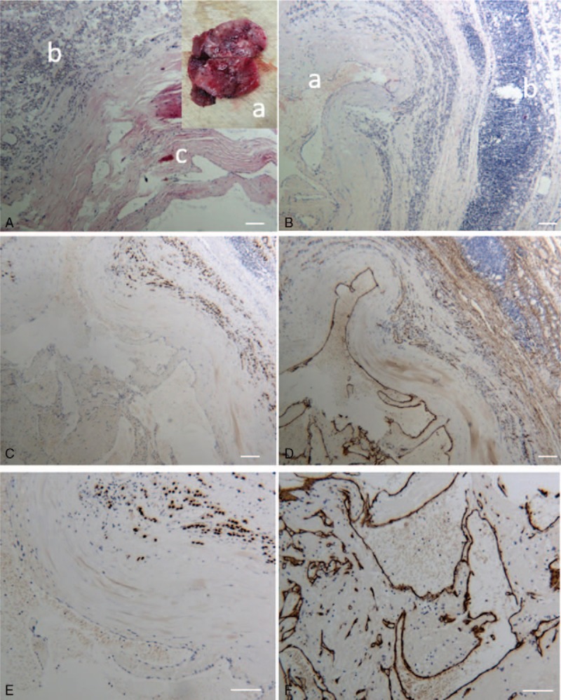
Diagnoses: Ultrasound and CT scan revealed a hypoechogenic and heterogeneous mass measuring 4 × 3.5 cm located in the right lobe of thyroid gland. Postoperative pathological and immunohistochemical examinations of the surgical specimen revealed a primary hemangioma of the thyroid gland.
Interventions: The patient received a right lobectomy of the thyroid.
Outcomes: The patient had been followed up for 10 months after surgery without complications and remained asymptomatic.
Lessons: Primary thyroid hemangioma should be considered when there is a well-circumscribed capsule mass on medical imaging without history of FNA or any other cervical procedures or trauma.
Conflict of interest statement
The authors report no conflicts of interest.
Figures
References
-
- Adams DM, Lucky AW. Cervicofacial vascular anomalies. I. Hemangiomas and other benign vascular tumors. Semin Pediatr Surg 2006;15:124–32. - PubMed
-
- Zara-Lopes T, Gimenez-Martins AP, Nascimento-Filho CH, et al. Role of MTHFR C677T and MTR A2756G polymorphisms in thyroid and breast cancer development. Genet Mol Res 2016;15:1–1. - PubMed
-
- Michalopoulos NV, Markogiannakis H, Kekis PB, et al. Primary cavernous hemangioma of the thyroid gland. South Med J 2010;103:674–5. - PubMed
-
- Gupta P. Primary cavernous hemangioma of the thyroid gland. South Med J 2013;103:674–5. - PubMed
-
- Gutzeit A, Stuckmann G, Tosoni I, et al. A cavernous hemangioma of the thyroid gland: first documentation by ultrasound of a rare pathology. J Clin Ultrasound 2011;39:172–4. - PubMed
Publication types
MeSH terms
LinkOut - more resources
Full Text Sources
Other Literature Sources
Medical
