. 2017 Oct 12;8(59):99261-99273.
doi: 10.18632/oncotarget.21818. eCollection 2017 Nov 21.

Constitutive Cyclin O deficiency results in penetrant hydrocephalus, impaired growth and infertility

Affiliations

Constitutive Cyclin O deficiency results in penetrant hydrocephalus, impaired growth and infertility

Marc Núnez-Ollé et al. Oncotarget. .

Abstract

Cyclin O (encoded by CCNO) is a member of the cyclin family with regulatory functions in ciliogenesis and apoptosis. Homozygous CCNO mutations have been identified in human patients with Reduced Generation of Multiple Motile Cilia (RGMC) and conditional inactivation of Ccno in the mouse recapitulates some of the pathologies associated with the human disease. These include defects in the development of motile cilia and hydrocephalus. To further investigate the functions of Ccno in vivo, we have generated a new mouse model characterized by the constitutive loss of Ccno in all tissues and followed a cohort during ageing. Ccno-/- mice were growth impaired and developed hydrocephalus with high penetrance. In addition, some Ccno+/- mice also developed hydrocephalus and affected Ccno-/- and Ccno+/- mice exhibited additional CNS defects including cortical thinning and hippocampal abnormalities. In addition to the CNS defects, both male and female Ccno-/- mice were infertile and female mice exhibited few motile cilia in the oviduct. Our results further establish CCNO as an important gene for normal development and suggest that heterozygous CCNO mutations could underlie hydrocephalus or diminished fertility in some human patients.

Keywords: Cyclin O; Gerotarget; ciliogenesis; development; hydrocephalus; neurogenesis.


Conflict of interest statement

CONFLICTS OF INTEREST The authors declare that they have no conflict of interest.

Figures
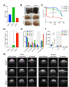
Figure 1
Figure 1. Ccno mutant mice develop hydrocephalus with high penetrance
A. Genotype distribution of the offspring derived from Ccno+/- matings (n=128). The distribution of genotypes differs from that expected by normal Mendelian inheritance ( p<0.0005, chi square test). B. Characteristic morphology of healthy Ccno+/+ and hydrocephalic Ccno-/- P23 littermates. The top and bottom view of the Ccno-/- brain shows generalized swelling due to the accumulation of cerebrospinal fluid. C. Kaplan-Meier survival plot of Ccno+/+, Ccno+/- and Ccno-/- mice. Littermates of mixed genotypes were kept in the same cage with free access to food and water and were sacrificed when moribund. The p-values were calculated using the log rank (Mantel-Cox) test. D. Incidence of hydrocephalus (percentage) in the cohort of mice used for the survival experiment (n=128; p<0.0005, one-way ANOVA with Bonferroni post hoc test). E. Pathologies found upon macroscopic examination in non-hydrocephalic mice (n=20 Ccno+/+, n=66 Ccno+/-, n=10 Ccno-/-). Differences among genotypes were not statistically significant by one-way ANOVA with Bonferroni post hoc test. F. Quantification of the volumes of the lateral (LV), third (3rd-V) and fourth (4th-V) brain ventricles of Ccno+/+ (n=9), Ccno+/- (n=11) and Ccno-/- (n=11) mice from the MRI images. Statistical analysis was done by one-way ANOVA with Tukey post hoc test. G. Representative examples of MRI images quantified in F. Young (upper left panels, mean age=53 days) and adult (upper right panels, mean age=160 days) Ccno+/+ (upper panels), Ccno+/- (middle panels) and Ccno-/- (lower panels) mice were analyzed. The location of the lateral (lv), third (3v) and fourth (4v) ventricles in the MRI images is indicated with purple arrowheads.
Figure 2
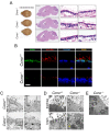
Figure 2. Loss of multiciliated cells in the ependymal and tracheal epithelia of Ccno-/- mice
A. Top view of the brain (left panels), sagittal sections (middle panels, H&E staining) and magnifications from the areas indicated by black rectangles (lateral and third ventricles, right panels, H&E staining) from Ccno+/+ (upper panels), Ccno+/- (middle panels) and Ccno-/- mice (lower panels). Scale bars = 2.5 mm (left panel) and 20 µm (right panels). B. Complete loss of immunoreactivity in Ccno-/- ependymal cells. Confocal microscopy images of formaldehyde-fixed, paraffin embedded sections of brain from Ccno+/+ (upper panels) and Ccno-/- (lower panels) adult mice were stained with antibodies against CCNO (green) and acetylated-α-Tubulin (red) and nuclei were counterstained with TO-PRO-3 (pseudocolored blue). Scale bar = 10 µm. C. Representative image of the ependymal epithelia from Ccno+/+ (upper panels) or Ccno-/- (lower panels) by TEM. Ciliated and non-ciliated cells are seen in Ccno+/+ sections, with cilia (c), basal bodies (bb) and microvilli (mv) becoming apparent in higher magnification views (square regions). In contrast, few or no cilia are seen in Ccno-/- cells and they are replaced by more abundant microvilli (mv). sc, secretory cells. Scale bars = 1 µm. D. Undocked, morphologically aberrant basal bodies (marked with asterisks) are seen in high magnification views of tracheal (upper panels) and brain (lower panels) sections of Ccno-/- mice. A frayed, greatly altered basal body in the ependyma of Ccno-/- mice is indicated with a yellow arrowhead. Scale bars = 500 nm. E. Accumulation of aggregates of modified microtubules are detected in tracheal sections of Ccno-/- mice, most likely corresponding to dysmorphic deuterosomes [8]. Scale bar = 500 nm.
Figure 3
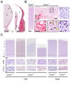
Figure 3. Neuronal damage in the CNS of Ccno-/- mice
A. Hydrocephalic Ccno-/- mice developed subdural hygroma. p: brain parenchyma; s: skull; m: meninges; c: cerebrospinal fluid; d: dural membrane; pi: pia mater. Scale bar = 200 µm. B. Intraparenchymal hemorrhage in a hydrocephalic P23 Ccno-/- mouse (central upper panel); in the left panel an image of the brain cortex of a P23 Ccno+/+ control mouse is shown. Bars = 200µm. Magnifications of the boxed areas containing the bleeding (b) and the neurons with pyknotic nuclei (p, yellow arrowheads) and vacuolated parenchyma (v, black arrowheads) are shown in the central lower panels. Scale bar = 100µm. The lower right panel shows acidophilic pyramidal neurons (black arrowheads) in the cortex of a P23 Ccno-/- mouse. The corresponding Ccno+/+ control is shown in the upper right panel. Scale bar = 20 µm. C. Cortical thinning in the brain of Ccno-/- mice. The layered structure present in Ccno+/+ and non-hydrocephalic P22 Ccno-/- mice (left image) is altered in hydrocephalic (middle image) and severely affected in highly hydrocephalic, moribund P22 Ccno-/- mice (right image). Acidophilic neurons and parenchyma vacuolation can be appreciated. The cortical layers are comparable in aged (P403) Ccno+/+ and Ccno-/- non-hydrocephalic littermates (right panels). Scale bar = 200 µm. A magnification of the neurons from the II and III layers is shown in the middle panels of the Figure. The innermost end of the cortical images containing the ependyma is shown in the lower panels. Scale bars = 20 µm. wm: White matter.
Figure 4
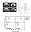
Figure 4. Hippocampal abnormalities in Ccno mutant mice
A. Representative MRI images of the hippocampi quantified in B. The areas delimited in yellow correspond to the hippocampal area quantified. c: brain cortex; lv: lateral ventricle. B. Quantification of the volumes of the hippocampi of Ccno+/+ (n=9), Ccno+/- (n=11) and Ccno-/- (n=11) mice from the MRI images. Statistical analysis was done by one-way ANOVA with the Tukey post hoc test. C. Correlation plots between the volume of the ventricles and the volume of the hippocampus quantified from the MRI images. A significant Pearson correlation test was obtained between the total volume of the ventricles or the volume of the lateral ventricles and the volume of the hippocampus.
Figure 5
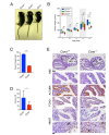
Figure 5. Impaired growth and infertility of Ccno mutant mice
A. Representative image of Ccno+/+, Ccno+/-, and Ccno-/- littermates at P30. B. Weights of Ccno+/+ (blue bars), Ccno+/- (green bars) and Ccno-/- mice (red bars) from 21 to 742 days postpartum. Age group 1: P21-P34; Age group2: P35-P49; Age group 3: P50-P99; Age group 4: P100-P499; Age group 5: P500-P742. Circles represent outliers. A minimum of five animals were plotted for each age group. Statistical significance was determined by the unpaired two-way Mann–Whitney U test. C and D. Quantification of the number of ciliated cells (C) and the number of cilia per cell (D). Statistical significance was determined by the unpaired Student’s t-test. E. Histochemical analysis of the oviductal epithelium reveals few MCCs but most of the cells of the epithelium expressing the MCC lineage marker FOXJ1 and are negative for MKi67 in young (P129) Ccno-/- mice. A positive control for MKi67 (granulosa cells from the ovary) are shown in the bottom panels. m: mucosa; am: ampulla; s: stroma; gr: granulosa; f: follicle. Scale bars = 100µm (top panels) and 20µm (rest of the panels).

References

    1. Lim S, Kaldis P. Cdks, cyclins and CKIs: roles beyond cell cycle regulation. Development. 2013;140:3079–3093. - PubMed
    1. Granes F, Roig MB, Brady HJ, Gil-Gomez G. Cdk2 activation acts upstream of the mitochondrion during glucocorticoid induced thymocyte apoptosis. Eur J Immunol. 2004;34:2781–2790. - PubMed
    1. Roig MB, Roset R, Ortet L, Balsiger NA, Anfosso A, Cabellos L, Garrido M, Alameda F, Brady HJ, Gil-Gomez G. Identification of a novel cyclin required for the intrinsic apoptosis pathway in lymphoid cells. Cell Death Differ. 2009;16:230–243. - PubMed
    1. Wallmeier J, Al-Mutairi DA, Chen CT, Loges NT, Pennekamp P, Menchen T, Ma L, Shamseldin HE, Olbrich H, Dougherty GW, Werner C, Alsabah BH, Kohler G, et al. Mutations in CCNO result in congenital mucociliary clearance disorder with reduced generation of multiple motile cilia. Nat Genet. 2014;46:646–651. - PubMed
    1. Amirav I, Wallmeier J, Loges NT, Menchen T, Pennekamp P, Mussaffi H, Abitbul R, Avital A, Bentur L, Dougherty GW, Nael E, Lavie M, Olbrich H, et al. Systematic Analysis of CCNO Variants in a Defined Population: Implications for Clinical Phenotype and Differential Diagnosis. Hum Mutat. 2016;37:396–405. - PubMed