. 2017 Sep 16;8(59):99825-99840.
doi: 10.18632/oncotarget.21076. eCollection 2017 Nov 21.

Saikosaponin-d, a calcium mobilizing agent, sensitizes chemoresistant ovarian cancer cells to cisplatin-induced apoptosis by facilitating mitochondrial fission and G2/M arrest

Affiliations

Saikosaponin-d, a calcium mobilizing agent, sensitizes chemoresistant ovarian cancer cells to cisplatin-induced apoptosis by facilitating mitochondrial fission and G2/M arrest

Hideaki Tsuyoshi et al. Oncotarget. .

Abstract

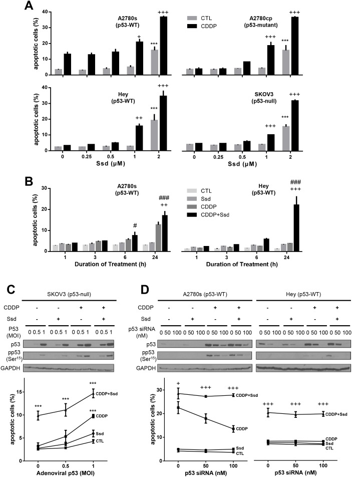
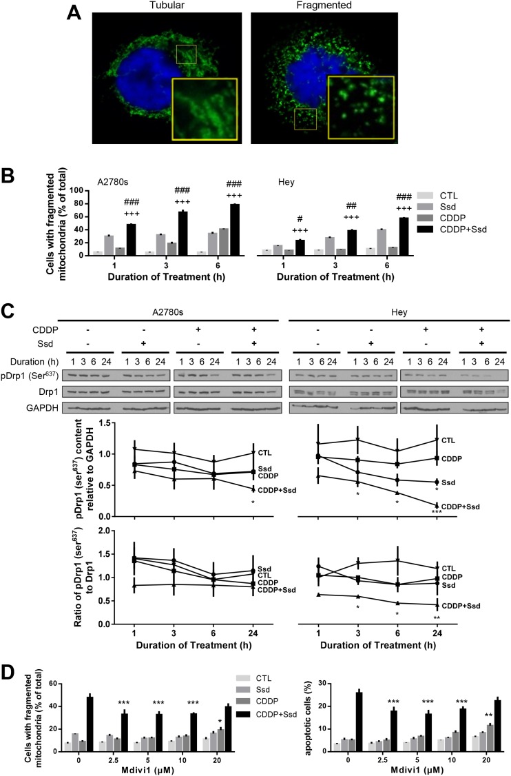
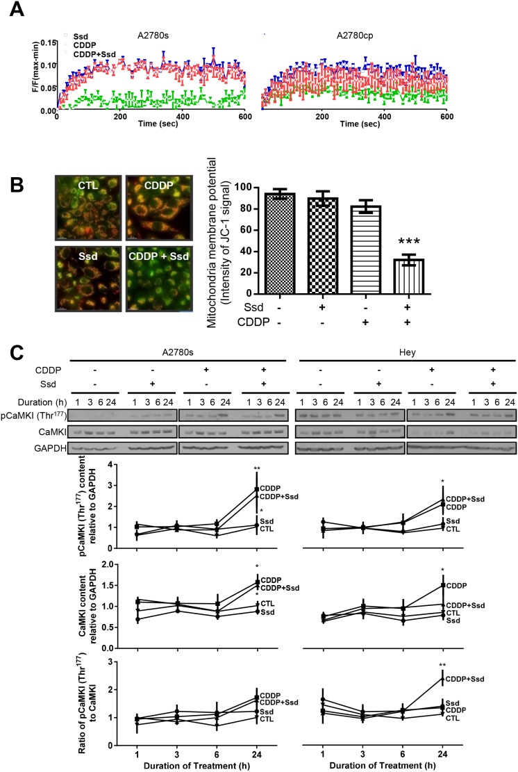
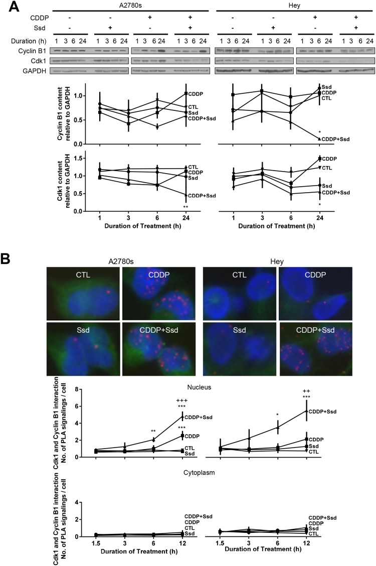
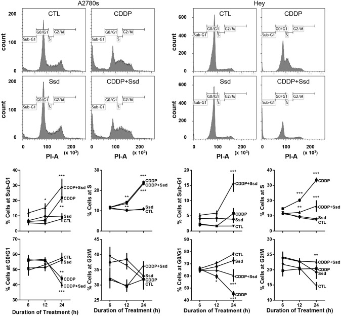
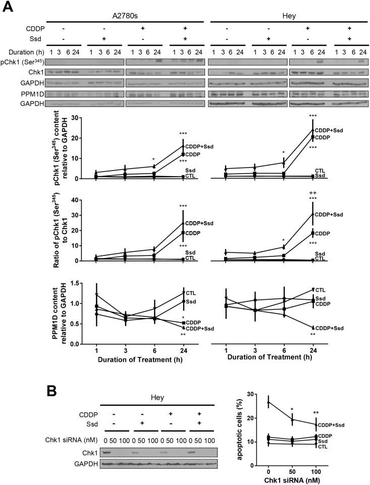
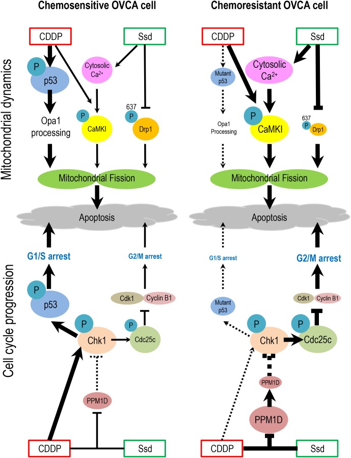
Cisplatin (CDDP) and its derivatives are first line anti-cancer drugs for ovarian cancer (OVCA). However, chemoresistance due to high incidence of p53 mutations leads to poor clinical prognosis. Saikosaponin-d (Ssd), a saponin from a herbal plant extract, has been shown to induce cell death and sensitize chemoresistant cells to chemotherapeutic agents. Here, we demonstrated that Ssd sensitized chemoresistant OVCA cells with either p53-wt, -mutant and -null to CDDP. The action of Ssd appears to be through induction of mitochondrial fragmentation and G2/M arrest. Ssd is mediated via calcium signaling, up-regulation of the mitochondrial fission proteins Dynamin-related protein 1 (Drp1) and optic atrophy 1 (Opa1), and loss in mitochondrial membrane potential (MMP). Moreover, in the presence of CDDP, Ssd also down-regulates protein phosphatase magnesium-dependent 1 D (PPM1D) and increases the phosphorylation of checkpoint protein kinases (Chk) 1, cell division cycle 25c (Cdc25c) and Cyclin dependent kinase 1 (Cdk1). Our findings suggest that Ssd could sensitize OVCA to CDDP independent of the p53 status through multiple signaling pathways. They support the notion that Ssd may be a novel adjuvant for the treatment of chemoresistant OVCA.

Keywords: G2/M arrest; Ssd; chemoresistance; mitochondrial dynamics; ovarian cancer.


Conflict of interest statement

CONFLICTS OF INTEREST The authors declare no conflicts of interest.

Figures

Figure 1
Figure 1. Ssd-induced apoptosis and sensitization of OVCA cells to CDDP in p53-independent manner
(A) Ssd increased apoptosis and in the presence of CDDP Ssd sensitized OVCA cells to CDDP regardless of p53 status. A2780s, A2780cp, Hey and SKOV3 cells were cultured with CDDP (10 μM) and/or Ssd (0-2 μM, 24 h) and apoptosis were assessed. (B) Ssd-induced sensitization was time-dependent. A2780s and Hey cells were cultured with CDDP (10 μM) and/or Ssd (1 μM, 0-24 h). (C) The infection with adenoviral p53 had no significant affect on Ssd-sensitized apoptosis. SKOV3 cells were infected with adenoviral p53 (MOI = 0–1; 4 h), cultured with CDDP (10 μM) and/or Ssd (1 μM, 24 h) and apoptosis were assessed. p53, anti-phospho-Ser15-p53 and GAPDH contents were assessed by WB. (D) The treatment with p53 siRNA failed to decrease Ssd-sensitized apoptosis. A2780s and Hey cells were treated with p53 siRNA (0–100 nM, 12 h), cultured with CDDP (10 μM) and/or Ssd (1 μM, 24 h). ***P<0.001 (vs respective CTL), +P<0.05, ++P<0.01 and +++P<0.001 (vs respective CDDP) and #P<0.05 and ###P<0.001 (vs respective Ssd). (n=3).
Figure 2
Figure 2. Ssd-induced mitochondria fragmentation via decrease of phospho-Ser637-Drp1 in chemoresistant OVCA cells
(A) OVCA cells showed two major mitochondrial phenotypes: tubular and fragmented. The inset is an enlarged image of box area. (B) Ssd induced mitochondrial fragmentation and sensitized chemoresistant OVCA cells to CDDP. A2780s and Hey cells were cultured with CDDP (10 μM) and/or Ssd (1 μM, 0-6 h) and mitochondrial phenotype was assessed (n=3). #P<0.05, ##P<0.01 and ###P<0.001 (vs respective Ssd) and +++P<0.001 (vs respective CDDP). (C) Ssd decreased phospho-Ser637-Drp1 content and the combination with CDDP decreased phospho-Ser637-Drp1 content and the ratio of phospho-Ser637-Drp1 to Drp1 more than Ssd alone. A2780s and Hey cells were cultured with CDDP (10 μM) and/or Ssd (1 μM, 0-24 h). Drp1, phospho-Ser637-Drp1 and GAPDH contents were assessed by WB (n=3).*P<0.05, **P<0.01 and***P<0.001 (vs respective CTL). (D) Ssd-induced mitochondrial fragmentation and apoptosis were attenuated by mdivi1. Hey cells were cultured with CDDP (10 μM), Ssd (1 μM) and mdivi1 (0-20 μM, 24 h). Mitochondrial phenotype and apoptosis were assessed (n=3). *P<0.05, **P<0.01 and ***P<0.001 (vs respective mdivi1 = 0 μM).
Figure 3
Figure 3. Ssd-increased [Ca2+]c and subsequent MMP loss and activation of CaMKI in chemoresistant OVCA cells
(A) Ssd increased [Ca2+]c in OVCA cells, but not CDDP. A2780s and A2780cp cells were cultured with CDDP (10 μM) and/or Ssd (2 μM) and [Ca2+]c was measured using the FLIPR Calcium 6 Assay Kit. (B) CDDP with Ssd caused MMP loss. A2780cp cells were cultured with CDDP (10 μM) and/or Ssd (2 μM, 24 h) and immunostained with JC-1. (C) CDDP in the presence of Ssd increased phospho-Thr177-CaMKI contents and the ratio of phospho-Thr177-CaMKI to CaMKI. A2780s and Hey cells were cultured with CDDP (10 μM) and/or Ssd (1 μM, 0-24 h). CaMKI, phospho-Thr177-CaMKI and GAPDH contents were assessed by WB.*P<0.05, **P<0.01 and ***P<0.001 (vs respective CTL). (n=3).
Figure 4
Figure 4. Ssd, in combination with CDDP, decreases mitosis and increases subsequent apoptosis via inhibition of Cdk1 and Cyclin B1 complex in OVCA cells
(A) CDDP with Ssd decreased Cdk1 and CyclinB1 contents. A2780s and Hey cells were cultured with CDDP (10μM) and/or Ssd (1μM, 0-24h). Cyclin B1, Cdk1 and GAPDH contents were assessed by WB. (B) CDDP with Ssd increased the interaction between Cdk1 and Cyclin B1 in chemoresistant OVCA cells. A2780s and Hey cells were cultured with CDDP (10 μM) and Ssd (1 μM, 0-12h) and the interaction between Cdk1 and Cyclin B1 was determined by PLA. *P<0.05, **P<0.01 and***P<0.001 (vs respective CTL) and ++P<0.01 and +++P<0.001 (vs respective CDDP). (n=3).
Figure 5
Figure 5. CDDP with Ssd induced G2/M arrest
A2780s and Hey cells were cultured with CDDP (10 μM) and/or Ssd (1 μM, 0-24 h), fixed and incubated with RNase A (1 mg/ml) and propidium iodide (25 μg/ml) and analyzed for cell cycle progression using flow cytometer (n=3).*P<0.05, **P<0.01 and***P<0.001 (vs respective CTL).
Figure 6
Figure 6. CDDP with Ssd-induced phosphorylation of Ser345-Chk1 via decrease of PPM1D, leading to G2/M arrest
(A) CDDP with Ssd increased phosphor-Ser345-Chk1 and decreased PPM1D contents. A2780s and Hey cells were cultured with CDDP (10 μM) and/or Ssd (1 μM, 0-24 h). Phosphor-Ser345-Chk1, Chk1, PPM1D and GAPDH contents were assessed by WB (n=3).*P<0.05, **P<0.01 and***P<0.001 (vs respective CTL) and ++P<0.01 (vs respective CDDP). (B) Chk1 siRNA attenuated Ssd-sensitized apoptosis. Hey cells were treated with Chk1 siRNA (0–100 nM, 12 h), cultured with CDDP (10 μM) and/or Ssd (1 μM, 24 h) and apoptosis were assessed (n=3). *P<0.05 and **P<0.01 (vs respective siRNA = 0 nM).
Figure 7
Figure 7. Hypothetical model illustrating the sensitization of chemoresistant OVCA cells by Ssd
In chemosensitive cells, CDDP treatment results in the phosphorylation and activation of p53 and activation of Oma1, leading to Opa1 processing, mitochondrial fission and apoptosis. CDDP also activates Chk1, which phosphorylates p53, leading to G1/S arrest and apoptosis. In chemoresistant cells which often exhibit high incidence of p53 mutation and increased PPM1D stability, which inhibits Chk1 activity, CDDP fails to induce G1/S arrest and Opa1 processing. However, Ssd suppresses phospho-Ser637-Drp1 content, increases [Ca2+]c and, in the presence of CDDP, decreases MMP and increases CaMKI phosphorylation. These actions of Ssd lead to mitochondrial fission and subsequent apoptosis. Moreover, in the presence of CDDP, Ssd decreases PPM1D level, activates Chk1 and increases phospho-Cdc25c content, resulting in G2/M arrest and apoptosis.

References

    1. Canadian Cancer Society's Advisory Committee on Cancer Statistics Canadian Cancer Statistics. 2016
    1. Olivier M, Hollstein M, Hainaut P. TP53 mutations in human cancers: origins, consequences, and clinical use. Cold Spring Harbor perspectives in biology. 2010;2:a001008. - PMC - PubMed
    1. Wong VK, Zhou H, Cheung SS, Li T, Liu L. Mechanistic study of saikosaponin-d (Ssd) on suppression of murine T lymphocyte activation. Journal of cellular biochemistry. 2009;107:303–315. - PubMed
    1. Wong VK, Zhang MM, Zhou H, Lam KY, Chan PL, Law CK, Yue PY, Liu L. Saikosaponin-d Enhances the Anticancer Potency of TNF-alpha via Overcoming Its Undesirable Response of Activating NF-Kappa B Signalling in Cancer Cells. Evidence-based complementary and alternative medicine. 2013;2013:745295. - PMC - PubMed
    1. Hsu YL, Kuo PL, Chiang LC, Lin CC. Involvement of p53, nuclear factor kappaB and Fas/Fas ligand in induction of apoptosis and cell cycle arrest by saikosaponin d in human hepatoma cell lines. Cancer letters. 2004;213:213–221. - PubMed