

MicroRNA-433 inhibits oral squamous cell carcinoma cells by targeting FAK
- PMID: 29245973
- PMCID: PMC5725015
- DOI: 10.18632/oncotarget.22151
MicroRNA-433 inhibits oral squamous cell carcinoma cells by targeting FAK
Retraction in
-
Retraction: MicroRNA-433 inhibits oral squamous cell carcinoma cells by targeting FAK.Oncotarget. 2022 Sep 14;13:1033. doi: 10.18632/oncotarget.28270. eCollection 2022. Oncotarget. 2022. PMID: 36128325 Free PMC article.
Abstract
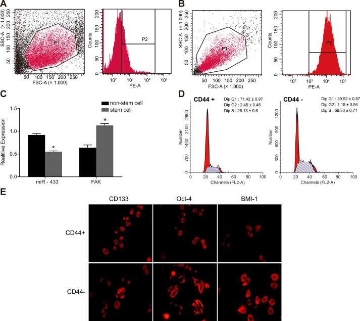
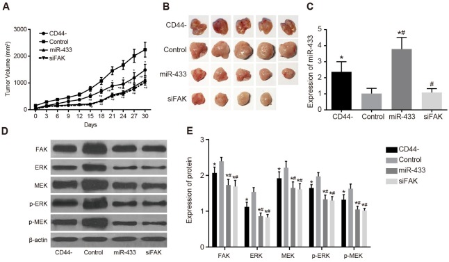
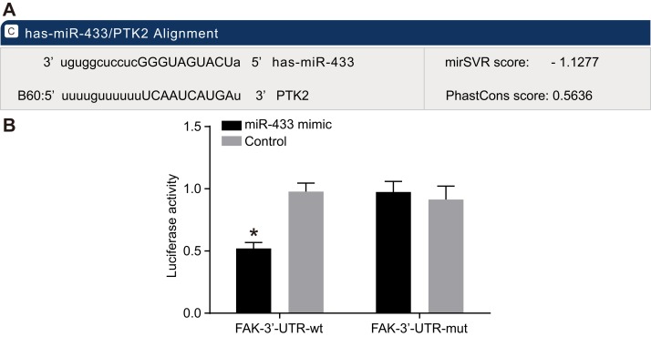
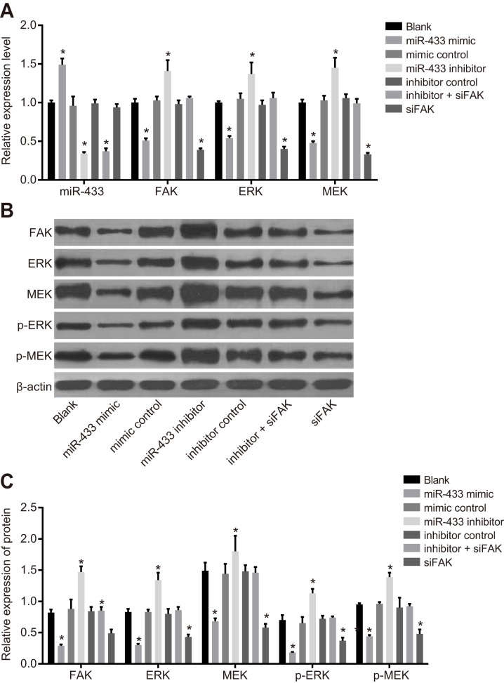
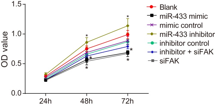
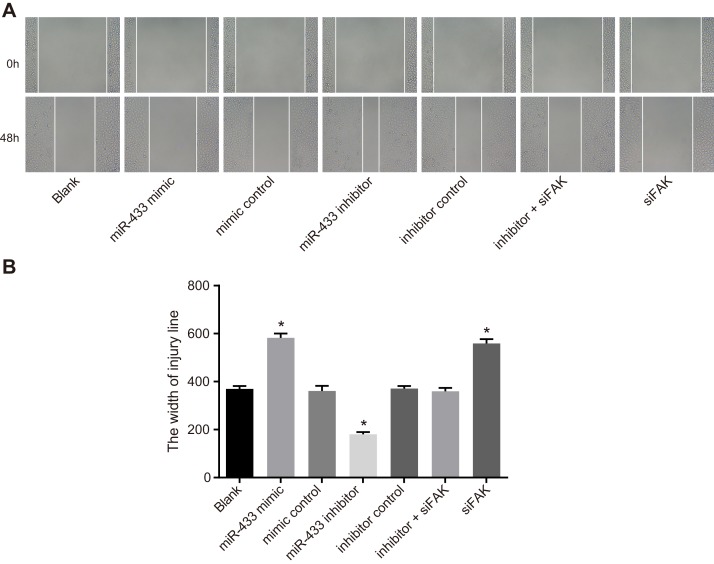
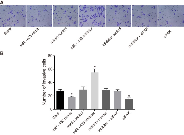
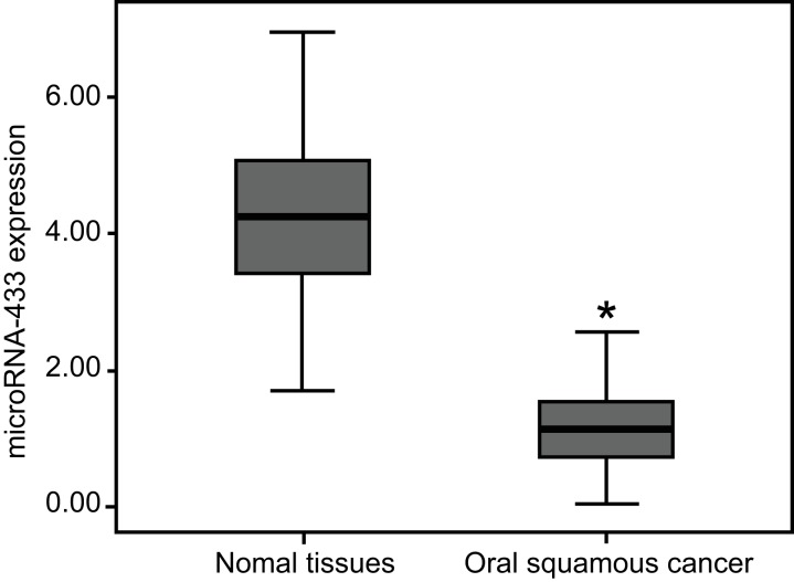
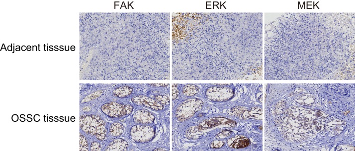
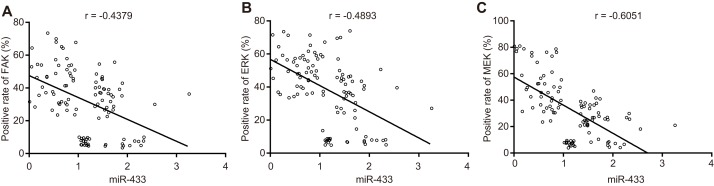
We investigated the involvement of microRNA-433 (miR-433) in the proliferation, migration, and invasiveness of oral squamous cell carcinoma (OSCC). Totally 108 OSCC tissues and adjacent normal tissues from patients with OSCC were collected. Also, transplanted tumor formation experiment in nude mice was conducted to verify the effect of miR-433 and FAK on subcutaneous transplanted tumor. The CD44+ stem cell from SCC-9 were collected and assigned into the blank, miR-433 mimics, mimics control, miR-433 inhibitors, inhibitors control, siFAK and miR-433 inhibitors + siFAK groups. The qRT-PCR and western blotting were used to detect miR-433, FAK, ERK, MEK, pERK and pMEK after transfection. Flow cytometry, MTT assay, scratch test and Transwell assay were performed to determine the cell proportion, growth, migration and invasion of SCC-9 cells. In cell line SCC-9, expression of CD133, Oct-4, and BIM-1 was greater in CD44+ cells than CD44- cells, indicating that CD44+ cells had characteristics of tumor stem cells. Expression of FAK, ERK, MEK, p-ERK and p-MEK was decreased in tumor tissues from the CD44-, miR-433, and siFAK groups. Expression of MiR-433 mRNA was elevated, while levels of FAK, ERK, MEK, p-ERK, and p-MEK mRNA were all decreased in the miR-433 mimics group. In the miR-433 mimics and siFAK groups, cell proliferation, migration, and invasion were all decreased, while the opposite trends were seen in the miR-433 inhibitor group. These results indicate that miR-433 downregulates FAK through the ERK/MAPK signaling pathway to inhibit the proliferation, migration, and invasiveness of SCC-9 OSCC cells.
Keywords: ERK; FAK; SCC-9 cell; microRNA-433; oral squamous cell carcinoma.
Conflict of interest statement
CONFLICTS OF INTEREST The authors declare no conflicts of interest.
Figures
References
-
- Chen D, Yan W, Liu Z, Zhang Z, Zhu L, Liu W, Ding X, Wang A, Chen Y. Downregulation of miR-221 enhances the sensitivity of human oral squamous cell carcinoma cells to Adriamycin through upregulation of TIMP3 expression. Biomed Pharmacother. 2016;77:72–8. - PubMed
-
- Choi S, Myers JN. Molecular pathogenesis of oral squamous cell carcinoma: implications for therapy. J Dent Res. 2008;87:14–32. - PubMed
-
- Peng CH, Liao CT, Ng KP, Tai AS, Peng SC, Yeh JP, Chen SJ, Tsao KC, Yen TC, Hsieh WP. Somatic copy number alterations detected by ultra-deep targeted sequencing predict prognosis in oral cavity squamous cell carcinoma. Oncotarget. 2015;6:19891–906. https://doi.org/10.18632/oncotarget.4336. - DOI - PMC - PubMed
-
- Sanghvi S, Khan MN, Patel NR, Yeldandi S, Baredes S, Eloy JA. Epidemiology of sinonasal squamous cell carcinoma: a comprehensive analysis of 4994 patients. Laryngoscope. 2014;124:76–83. - PubMed
-
- Lin CC, Chen PC, Lein MY, Tsao CW, Huang CC, Wang SW, Tang CH, Tung KC. WISP-1 promotes VEGF-C-dependent lymphangiogenesis by inhibiting miR-300 in human oral squamous cell carcinoma cells. Oncotarget. 2016;7:9993–10005. https://doi.org/10.18632/oncotarget.7014. - DOI - PMC - PubMed
Publication types
LinkOut - more resources
Full Text Sources
Other Literature Sources
Research Materials
Miscellaneous
