

Individualized coracoid osteotomy and 3D congruent arc reconstruction of glenoid for the treatment of recurrent anterior shoulder dislocation
- PMID: 29246239
- PMCID: PMC5732511
- DOI: 10.1186/s13018-017-0687-8
Individualized coracoid osteotomy and 3D congruent arc reconstruction of glenoid for the treatment of recurrent anterior shoulder dislocation
Abstract
Background: The present study investigated individualized coracoid osteotomy for 3D congruent arc glenoid reconstruction and evaluated the clinical outcomes in recurrent anterior shoulder dislocation.
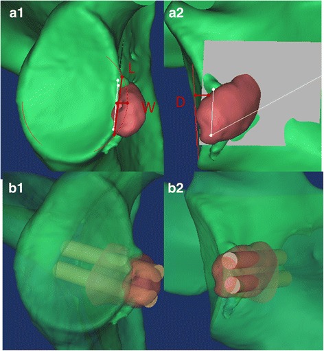
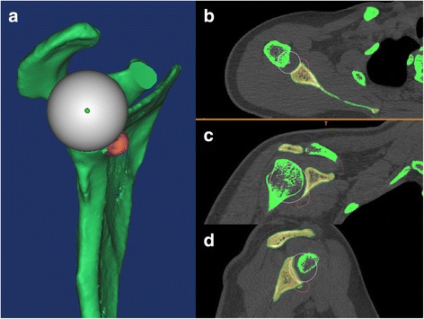
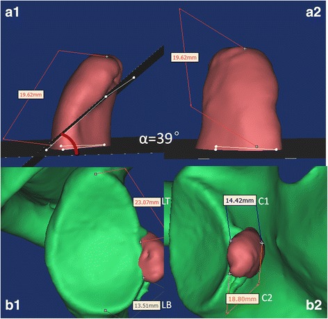
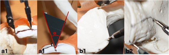
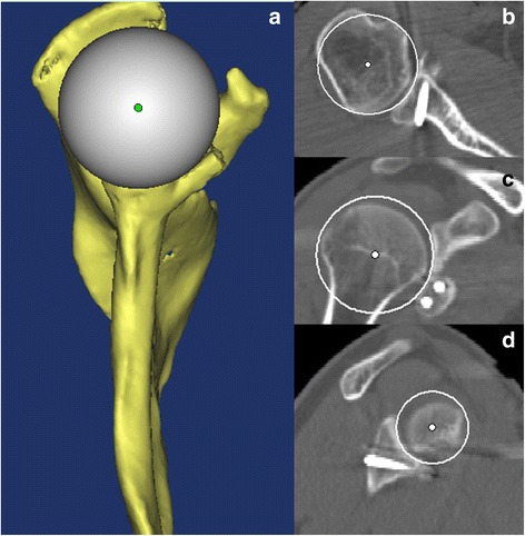
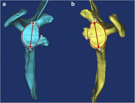
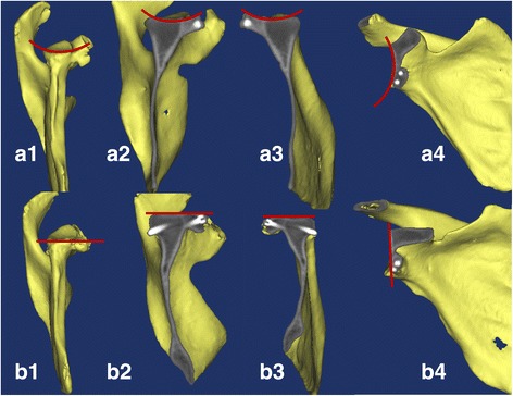
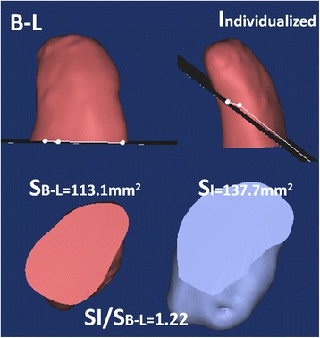
Methods: From January 2005 to July 2015, 78 patients with glenoid defect underwent coracoid and conjoint tendon transposition. The patients were divided into the individualized group (n = 34) and the non-individualized group (n = 44). All patients had CT data to reconstruct the shoulder model using Mimics software. In the individualized group, the individual coracoid osteotomy and bone fixation position parameters were measured from preoperative planification through simulating a 3D congruent arc glenoid reconstruction model. The non-individualized group underwent classic Bristow-Latarjet (B-L) procedure. The postoperative evaluation parameters included 3D congruent arc index, coracoid bone position, shoulder osteoarthritis index (Samilson-Prieto) and shoulder function score (Rowe, Constant-Murley score).
Results: The mean follow-up time was 51.0 months (ranging from 24 to 146). The individualized group got 3D congruent arc reconstruction of the glenoid by postoperative CT scanning. Bone position was more precise in the individual group than that in the B-L group. There was a lower incidence of shoulder osteoarthritis (Samilson-Prieto) in the individual group compared with that in the B-L group: 0 vs 13.6% (mild 6/44, P = 0.027), respectively. No significant difference was observed between the individual and B-L groups in rate of re-dislocation: 0 vs 4.5% (2/44, P = 0.315), respectively. The postoperative Rowe and Constant score was significantly improved but was not significantly different between the two groups.
Conclusion: The individual procedure achieved 3D congruent arc glenoid reconstruction. The clinical effects in patients with medium glenoid defect were good, especially the low incidence of shoulder osteoarthritis in middle-term follow-up.
Keywords: Bone defect; Congruent arc; Coracoid transposition; Preoperative planning; Shoulder dislocation; Three-dimensional reconstruction.
Conflict of interest statement
Ethics approval and consent to participate
The study was approved by the Clinical Academic Committee of the Third Military Medical University Southwest Hospital and was approved by all the members. The study was conducted in compliance with the Helsinki Declaration.
Consent for publication
All patients gave written, informed consent for publication.
Competing interests
The authors declare that they have no competing interests.
Publisher’s Note
Springer Nature remains neutral with regard to jurisdictional claims in published maps and institutional affiliations.
Figures
Similar articles
-
[Application of LU-tarjet congruent-arc technique in treatment of recurrent shoulder dislocation with huge glenoid defect].Zhongguo Xiu Fu Chong Jian Wai Ke Za Zhi. 2024 Jun 15;38(6):672-678. doi: 10.7507/1002-1892.202403039. Zhongguo Xiu Fu Chong Jian Wai Ke Za Zhi. 2024. PMID: 38918186 Free PMC article. Chinese.
-
[Analysis of the effectiveness of coracoid osteotomy and concentric coaxial reconstruction of the glenoid cavity in the treatment of recurrent anterior dislocation of the shoulder joint].Zhonghua Yi Xue Za Zhi. 2022 Aug 9;102(29):2283-2289. doi: 10.3760/cma.j.cn112137-20211121-02593. Zhonghua Yi Xue Za Zhi. 2022. PMID: 35927060 Chinese.
-
[Early-term effectiveness of Latarjet procedure by coracoid osteotomy with preserving coracoacromial ligament for recurrent anterior shoulder dislocation].Zhongguo Xiu Fu Chong Jian Wai Ke Za Zhi. 2024 Jun 15;38(6):655-659. doi: 10.7507/1002-1892.202403108. Zhongguo Xiu Fu Chong Jian Wai Ke Za Zhi. 2024. PMID: 38918183 Free PMC article. Chinese.
-
Use of allograft to reconstruct anterior bony glenoid defect in chronic glenohumeral instability: a systematic review.Arch Orthop Trauma Surg. 2020 Oct;140(10):1475-1485. doi: 10.1007/s00402-020-03511-6. Epub 2020 Jun 10. Arch Orthop Trauma Surg. 2020. PMID: 32524228
-
Comparative Systematic Review of Fixation Methods of the Coracoid and Conjoined Tendon in the Anterior Glenoid to Treat Anterior Shoulder Instability.Orthop J Sports Med. 2019 Jan 25;7(1):2325967118820539. doi: 10.1177/2325967118820539. eCollection 2019 Jan. Orthop J Sports Med. 2019. PMID: 30719477 Free PMC article. Review.
Cited by
-
To further incorporate computer-aided designs to improve preoperative planning in total hip arthroplasty: a cohort study.Front Surg. 2024 Jul 8;11:1345261. doi: 10.3389/fsurg.2024.1345261. eCollection 2024. Front Surg. 2024. PMID: 39040681 Free PMC article.
-
A ligament tensor-guided extramedullary alignment technique for distal femoral cut in total knee replacement: results at a minimum 3 years follow-up.Arch Orthop Trauma Surg. 2021 Dec;141(12):2295-2302. doi: 10.1007/s00402-021-04115-4. Epub 2021 Aug 12. Arch Orthop Trauma Surg. 2021. PMID: 34386837
References
MeSH terms
LinkOut - more resources
Full Text Sources
Other Literature Sources
Medical
