. 2017 Dec 16;14(1):249.
doi: 10.1186/s12974-017-1022-3.

Macrophages in trigeminal ganglion contribute to ectopic mechanical hypersensitivity following inferior alveolar nerve injury in rats

Affiliations

Macrophages in trigeminal ganglion contribute to ectopic mechanical hypersensitivity following inferior alveolar nerve injury in rats

Dulguun Batbold et al. J Neuroinflammation. .

Abstract

Background: Accidental mandibular nerve injury may occur during tooth extraction or implant procedures, causing ectopic orofacial pain. The exact mechanisms underlying ectopic orofacial pain following mandibular nerve injury is still unknown. Here, we investigated the role of macrophages and tumor necrosis factor alpha (TNFα) in the trigeminal ganglion (TG) in ectopic orofacial pain following inferior alveolar nerve transection (IANX).

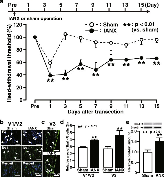
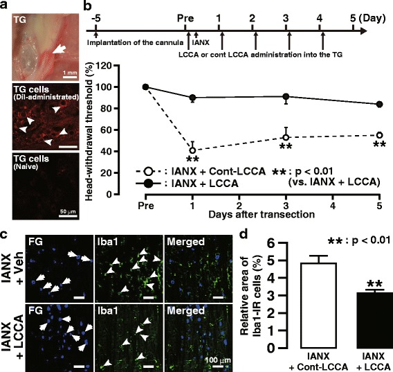
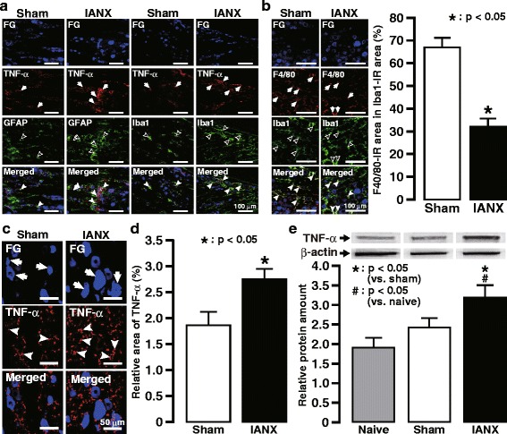
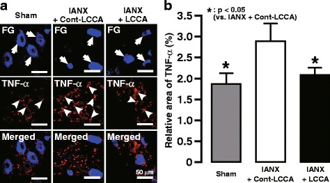
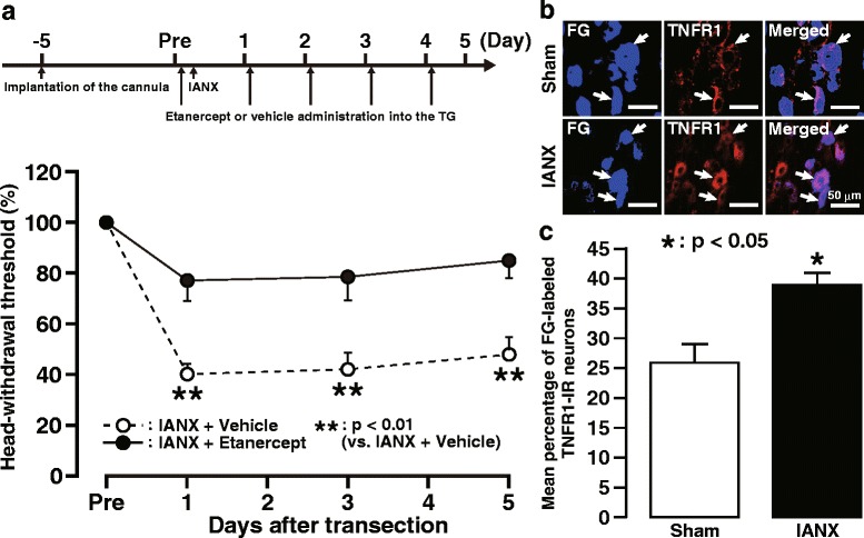
Methods: IANX was performed and the mechanical head-withdrawal threshold (MHWT) in the whisker pad skin ipsilateral to IANX was measured for 15 days. Expression of Iba1 in the TG was examined on day 3 after IANX, and the MHWT in the whisker pad skin ipsilateral to IANX was measured following successive intra-ganglion administration of the macrophage depletion agent liposomal clodronate Clophosome-A (LCCA). TNFα expression in the TG and the MHWT in the whisker pad skin ipsilateral to IANX following successive intra-ganglion administration of the TNFα blocker etanercept were measured on day 3 after IANX, and tumor necrosis factor receptor-1 (TNFR1) immunoreactive (IR) cells in the TG were analyzed immunohistochemically on day 3.

Results: The MHWT in the whisker pad skin was significantly decreased for 15 days, and the number of Iba1-IR cells was significantly increased in the TG on day 3 after IANX. Successive intra-ganglion administration of the macrophage depletion agent LCCA significantly reduced the increased number of Iba1-IR cells in the TG and reversed the IANX-induced decrease in MHWT in the whisker pad skin. TNFα expression was increased in the TG on day 3 after IANX and was reduced following successive intra-ganglion administration of the TNFα inhibitor etanercept. The decreased MHWT was also recovered by etanercept administration, and TNFR1-IR cells in the TG were increased on day 3 following IANX.

Conclusions: These findings suggest that signaling cascades resulting from the production of TNFα by infiltrated macrophages in the TG contributes to the development of ectopic mechanical allodynia in whisker pad skin following IANX.

Keywords: Ectopic orofacial pain; Inferior alveolar nerve transection; Macrophage; Mechanical allodynia; Trigeminal ganglion; Tumor necrosis factor alpha.


Conflict of interest statement

Ethics approval

The experimental protocols including all the surgical procedures and animal usages conformed to the guidelines for the care and use of laboratory animals by the National Institutes of Health Guide for the Care and Use of Laboratory Animals and the guidelines of the International Association for the Study of Pain. This study was approved by the Animal Experimentation Committee at Nihon University (AP16D010).

Consent for publication

Not applicable.

Competing interests

The authors declare that they have no competing interests.

Publisher’s Note

Springer Nature remains neutral with regard to jurisdictional claims in published maps and institutional affiliations.

Figures

Fig. 1
Fig. 1
Mechanical allodynia and Iba1 expression in the TG following IANX. a The timing of IANX or sham operation (upper) and the changes in MHWT of the ipsilateral whisker pad skin for 15 days following IANX or sham operation (lower). The MHWT was normalized to the pre-IANX MHWT (100%). Error bars indicate SEM. **p < 0.01 vs. sham (n = 6 in each; two-way ANOVA with repeated measures followed by Bonferroni’s multiple-comparison test). Iba1-IR cells in V1/V2 (b) and V3 (c) in the ipsilateral TG on day 3 following IANX or sham operation. Arrows indicate FG-labeled neurons. Arrowheads indicate Iba1-IR cells. d The relative area of Iba1-IR cells in V1/V2 and V3 in TG on day 3 following IANX and sham operation. Error bars indicate SEM. **p < 0.01 vs. sham. (n = 5 in each, Student’s t test). e The relative amount of Iba1 protein in TG on day 3 following IANX or sham operation. Error bars indicate SEM. *p < 0.05 vs. sham (n = 10 in each, Student’s t test)
Fig. 2
Fig. 2
Effect of intra-TG LCCA administration on mechanical allodynia and Iba1 expression following IANX. a Photograph (upper) and photomicrograph (middle) of TG on day 3 following DiI administration into the TG through the guide cannula and photomicrograph (lower) of TG in naive rat. Arrows indicate DiI labeling. b The timing of the cannula implantation, LCCA, or Cont-LCCA administration into the TG, IANX, or sham operation (upper) and the changes in MHWT of the ipsilateral whisker pad skin following daily intra-TG LCCA or Cont-LCCA administration following IANX (lower). The MHWT was normalized to the pre-IANX MHWT (100%). Arrows indicate the timing of intra-TG LCCA or Cont-LCCA administration. Error bars indicate SEM. **p < 0.01 vs. sham (n = 6 in each; two-way ANOVA with repeated measures followed by Bonferroni’s multiple-comparison test). c Iba1-IR cells and FG-labeled TG neurons in V1/V2 on day 3 following intra-TG LCCA or Cont-LCCA administration in IANX rats. Arrows indicate FG-labeled neurons. Arrowheads indicate Iba1-IR cells. d The relative area of Iba1-IR cells in V1/V2 in TG on day 3 following intra-TG LCCA or Cont-LCCA administration in IANX rats. Error bars indicate SEM. **p < 0.01 vs. sham. (n = 5 in each, Student’s t test)
Fig. 3
Fig. 3
TNFα expression in TG following IANX. a TNFα expression in the Iba1-IR cells or the GFAP-IR cells in ipsilateral TG on day 3 following IANX or sham operation. Arrows indicate TNFα-IR cells, open arrowheads indicate GFAP-IR cells or Iba1-IR cells, and close arrowheads indicate TNFα- and GFAP-IR cells or TNFα- and Iba1-IR cells. b F4/80 expression in the Iba1-IR cells in ipsilateral TG on day 3 following IANX or sham operation (left). Arrows indicate F4/80 expression in Iba1-IR cells, open arrowheads indicate Iba1-IR cells, and close arrowheads indicate F4/80- and Iba1-IR cells. The relative area of F4/80 immuno-products in Iba1-IR cells in the TG on day 3 following IANX or sham operation (right). Error bars indicate SEM. ***p < 0.001 vs. sham. (n = 5 in each, Student’s t test). c TNFα expression in ipsilateral TG on day 3 following IANX or sham operation. Arrows indicate FG-labeled neurons. Arrowheads indicate TNFα immuno-products. d The relative area of TNFα immuno-products in the TG on day 3 following IANX or sham operation. Error bars indicate SEM. *p < 0.05 vs. sham (n = 5 in each, Student’s t test). e The relative amount of TNFα protein in TG in naive animals or on day 3 following IANX or sham operation. Error bars indicate SEM. *p < 0.05 vs. sham. #p < 0.05 vs. naive (n = 5 in each, one-way ANOVA with Tukey’s multiple-comparison test)
Fig. 4
Fig. 4
Changes in TNFα expression in TG following LCCA administration following IANX. a TNFα expressed in V1/V2 in the TG on day 3 following intra-TG LCCA or Cont-LCCA administration in IANX rats or in sham-operated rats. Arrows indicate FG-labeled neurons. Arrowheads indicate TNFα immuno-products. b Changes in the relative area of TNF-α immuno-products in the TG on day 3 following intra-TG LCCA or Cont-LCCA administration in IANX rats or in sham-operated rats. Error bars indicate SEM. *p < 0.05 vs. Cont-LCCA. (n = 5 in each, one-way ANOVA with Tukey’s multiple-comparison test)
Fig. 5
Fig. 5
Effect of intra-TG etanercept administration on mechanical allodynia following IANX. a The timing of the cannula implantation, etanercept, or vehicle administration into the TG, IANX, or sham operation (upper) and changes in MHWT of the ipsilateral whisker pad skin by daily etanercept administration into TG following IANX (lower). The MHWT was normalized to the pre-IANX MHWT (100%). Arrows indicate the timing of intra-TG etanercept or vehicle administration. Error bars indicate SEM. **p < 0.01 vs. IANX + vehicle (n = 6 in each; two-way ANOVA with repeated measures followed by Bonferroni’s multiple-comparison test). b TNFR1 immunoreactivity in ipsilateral TG on day 3 following IANX or in sham-operated rats. Arrows indicate FG-labeled TNFR1-IR neurons. c Changes in the mean percentage of FG-labeled TNFR1-IR neurons on day 3 following IANX. Error bars indicate SEM. *p < 0.05 vs. sham (n = 5 in each, Student’s t test)

Similar articles

Cited by

References

    1. Renton T, Yilmaz Z, Gaballah K. Evaluation of trigeminal nerve injuries in relation to third molar surgery in a prospective patient cohort. Recommendations for prevention. Int J Oral Maxillofac Surg. 2012;41:1509–1518. doi: 10.1016/j.ijom.2012.06.025. - DOI - PubMed
    1. Khawaja N, Renton T. Case studies on implant removal influencing the resolution of inferior alveolar nerve injury. Br Dent J. 2009;206:365–370. doi: 10.1038/sj.bdj.2009.258. - DOI - PubMed
    1. Lin CS, SY W, Huang HY, et al. Systematic review and meta-analysis on incidence of altered sensation of mandibular implant surgery. PLoS One. 2016;11:e0154082. doi: 10.1371/journal.pone.0154082. - DOI - PMC - PubMed
    1. Kaji K, Shinoda M, Honda K, et al. Connexin 43 contributes to ectopic orofacial pain following inferior alveolar nerve injury. Mol Pain. 2016;12: doi:10.1177/1744806916633704. - PMC - PubMed
    1. Sugiyama T, Shinoda M, Watase T, et al. Nitric oxide signaling contributes to ectopic orofacial neuropathic pain. J Dent Res. 2013;92:1113–1117. doi: 10.1177/0022034513509280. - DOI - PubMed

Substances

LinkOut - more resources