. 2017 Dec 15:9:12-21.
doi: 10.1016/j.omtn.2017.08.007. Epub 2017 Aug 16.

Aptamer-Targeted Plasmonic Photothermal Therapy of Cancer

Affiliations

Aptamer-Targeted Plasmonic Photothermal Therapy of Cancer

Olga S Kolovskaya et al. Mol Ther Nucleic Acids. .

Abstract

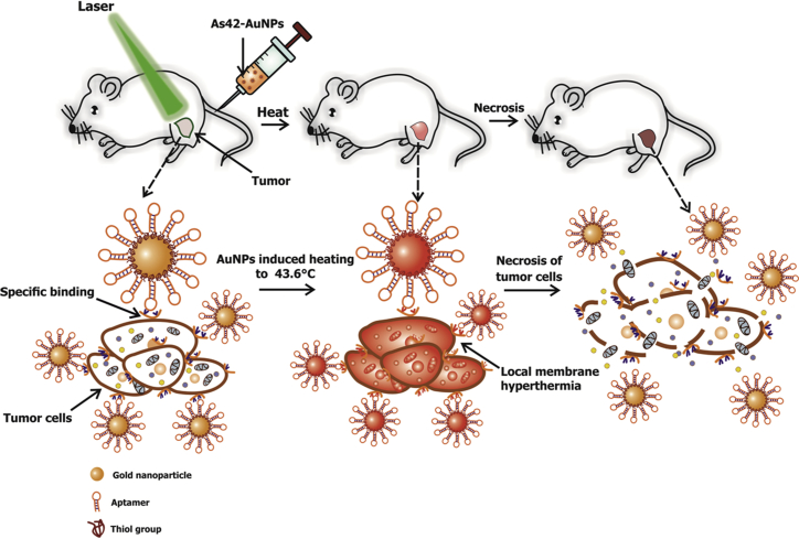
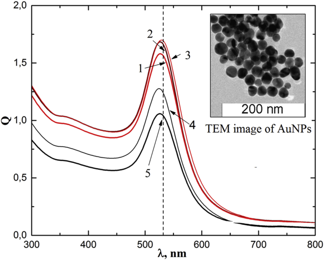
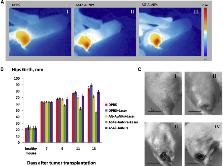
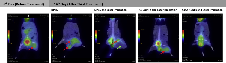
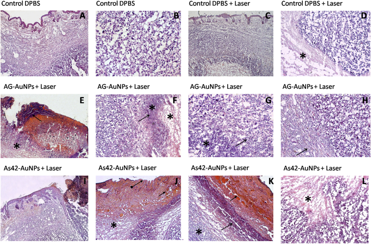
Novel nanoscale bioconjugates combining unique plasmonic photothermal properties of gold nanoparticles (AuNPs) with targeted delivery using cell-specific DNA aptamers have a tremendous potential for medical diagnostics and therapy of many cell-based diseases. In this study, we demonstrate the high anti-cancer activity of aptamer-conjugated, 37-nm spherical gold nanoparticles toward Ehrlich carcinoma in tumor-bearing mice after photothermal treatment. The synthetic anti-tumor aptamers bring the nanoparticles precisely to the desired cells and selectively eliminate cancer cells after the subsequent laser treatment. To prove tumor eradication, we used positron emission tomography (PET) utilizing radioactive glucose and computer tomography, followed by histological analysis of cancer tissue. Three injections of aptamer-conjugated AuNPs and 5 min of laser irradiations are enough to make the tumor undetectable by PET. Histological analysis proves PET results and shows lower damage of healthy tissue in addition to a higher treatment efficiency and selectivity of the gold nanoparticles functionalized with aptamers in comparison to control experiments using free unconjugated nanoparticles.

Keywords: DNA aptamer; computer tomography; gold nanoparticle; hyperthermia; mouse Ehrlich carcinoma; plasmonic photothermal therapy; positron emission tomography.


Figures

Figure 1
Figure 1
Scheme of Selective Elimination of Cancer Cells In Vivo Using As42-AuNPs in Plasmonic Photothermal Therapy As42-AuNPs are localized on the tumor cells after injection into a mouse tail vein. Local irradiation of a tumor site with a green laser causes nanoparticle heating and cell death followed by tumor eradication.
Figure 2
Figure 2
Absorption Spectra of Colloidal Solutions of AuNPs Curves 1–3 correspond to the following concentrations: 1.1 ⋅ 1012 mL−1 AuNPs; 4 − 1.1 ⋅ 109 mL−1 AuNPs; and 5 − 1.1 ⋅ 108 mL-1 AuNPs, respectively. Dashed line indicates the laser wavelength of 532 nm. Insert: transmission electron microscopy (TEM) image of AuNPs.
Figure 3
Figure 3
Effects of Photothermal Therapy of Ehrlich Carcinoma Cells Depending on the Presence of Gold Nanoparticles and/or DNA Aptamers In Vitro (A) Portions of dead cells were measured using trypan blue 3 hr after treatment in different experimental models. 1, intact Ehrlich carcinoma cells; 2, Ehrlich carcinoma cells after a 4-min laser irradiation; 3, Ehrlich carcinoma cells incubated with As42-AuNPs; 4, Ehrlich carcinoma cells preincubated with As42-AuNPs after 4 min of irradiation; 5, Ehrlich carcinoma cells incubated with free aptamer As42; 6, Ehrlich carcinoma cells incubated with free aptamer As42 after 10 min of irradiation; 7, Ehrlich carcinoma cells incubated with AG-AuNPs; 8, Ehrlich carcinoma cells incubated with AG-AuNPs after 4 min of irradiation. (B) Viability of Ehrlich cells after plasmonic photothermal therapy in vitro with As42-AuNPs (in the ratios of 10, 50, 100, and 200 AuNPs per cell). (C) Viability of liver and blood cell mixture after plasmonic photothermal therapy in vitro with As42-AuNPs (in the ratios of 10, 50, 100, and 200 AuNPs per cell). PI, propidium iodide. (D) Schematic representation of the Ehrlich, liver, and blood cell viability measurements after plasmonic photothermal treatment. All data are presented as the mean ± SEM.
Figure 4
Figure 4
Selectivity of Plasmonic Photothermal Therapy In Vitro (A) Necrosis in (1) intact Ehrlich carcinoma cells; (2) a liver and blood cell mixture (flow cytometry density plots); and (3) a mixture of Ehrlich carcinoma, liver and blood cells (flow cytometry dot plot). (B) Necrosis in (1) Ehrlich carcinoma cells; (2) a liver and blood cell mixture; and (3) a mixtue of Ehrlich carcinoma, liver, and blood cells (flow cytometry dot plots) after plasmonic photothermal therapy. (C) Schematic representation of (C1) necrotic Ehrlich carcinoma cells; (C2) intact liver and blood cells; and (C3) a mixture of Ehrlich carcinoma, liver, and blood cells after photothermal treatment.
Figure 5
Figure 5
Targeted Plasmonic Photothermal Therapy In Vivo (A) Thermal images of mouse hips after tail-vein injection of DPBS (AI), AG-AuNPs (AII) modified, or AS42-AuNPs (AIII) after 5 min of laser irradiation at 1.2 W. (B) Changes in the hip girth within tumors. The treatment has been performed on days 7, 9, and 11. (C) The representative images of the tumors of treated and non-treated mice are on day 11 treated with (I) DPBS only; (II) DPBS and 5 min of laser irradiation, (III) AG-AuNPs and 5 min of laser irradiation (IV) AS42-AuNPs and 5 min of laser irradiation. All data are presented as the mean ± SEM.
Figure 6
Figure 6
PET/CT and Histopathological Images of Mice after PPT Treatments (A–E) PET/CT images of mice before PPT treatment (A); after treatment with DPBS without laser irradiation (B); after PPT therapy with DPBS (C), AG-AuNPs (D), and AS42-AuNPs (E). Red arrows indicate accumulation of 18[F]-fluorodeoxyglucose; green arrow indicate necrosis and swelling.
Figure 7
Figure 7
Histopathological Assessment of Solid Ehrlich Carcinoma Tumor H&E staining. Representative sections were obtained after PPT with DPBS only. (A) Carcinoma under epidermis. (B) General view of mouse carcinoma treated with DPBS only. Magnification, 200×. PPT with DPBS and 5 min of laser irradiation. (C) General view of the tumor under epidermis. (D) The border between relatively intact viable and necrotic (asterisk) tumor tissues lacking inflammatory cells. Magnification, 100×. PPT with AG-AuNPs and 5 min of laser irradiation. (E) Ulcerative defect. (F) Scab (arrow) in the bottom of the wound and tumor necrosis under the dermis (asterisk). Magnification, 50×. (G) Complete destruction of tumor tissue in the center of the necrotic area (asterisk). Mostly dead segmented leukocytes (arrow). Magnification, ×100. (H) Inflammatory infiltration of segmented leukocytes at the tumor border (arrow). Magnification, 100×. PPT with As42-AuNPs and 5 min of laser irradiation. (I) Ulcerative defect, carcinoma necrosis (asterisk) in the bottom under the wound. (J) Necrosis of the skin and underlying tumor (asterisk), the loss of the epidermis (arrow), and the dermis bleeding (arrows). Magnification, 50×. (K) The boundary of the tumor necrosis in the dermis is separated with the area of leukocyte infiltration (arrow), outside of which hemocirculatory disorder takes place. Magnification, 50×. (L) Tumor necrosis (asterisk) is characterized by karyopyknosis, karyorhexis, and autolysis. Magnification, 100×.

References

    1. Lapotko D.O., Lukianova E., Oraevsky A.A. Selective laser nano-thermolysis of human leukemia cells with microbubbles generated around clusters of gold nanoparticles. Lasers Surg. Med. 2006;38:631–642. - PubMed
    1. Lavik E., von Recum H. The role of nanomaterials in translational medicine. ACS Nano. 2011;5:3419–3424. - PMC - PubMed
    1. Zharov V.P., Galitovskaya E.N., Johnson C., Kelly T. Synergistic enhancement of selective nanophotothermolysis with gold nanoclusters: potential for cancer therapy. Lasers Surg. Med. 2005;37:219–226. - PubMed
    1. Shi H., Ye X., He X., Wang K., Cui W., He D., II, Li D., Jia X. Au@Ag/Au nanoparticles assembled with activatable aptamer probes as smart “nano-doctors” for image-guided cancer thermotherapy. Nanoscale. 2014;6:8754–8761. - PubMed
    1. Kolovskaya O.S., Zamay T.N., Zamay A.S., Glazyrin Y.E., Spivak E.A., Zubkova O.A., Kadkina A.V., Erkaev E.N., Zamay G.S., Savitskaya A.G. DNA-aptamer/protein interaction as a cause of apoptosis and arrest of proliferation in Ehrlich ascites carcinoma cells. Biochemistry (Mosc). Suppl. Ser. A Membr. Cell Biol. 2014;8(1):60–72.

LinkOut - more resources