. 2017 Dec 15:9:170-181.
doi: 10.1016/j.omtn.2017.09.004. Epub 2017 Sep 14.

Antisense Oligonucleotides Targeting Y-Box Binding Protein-1 Inhibit Tumor Angiogenesis by Downregulating Bcl-xL-VEGFR2/-Tie Axes

Affiliations

Antisense Oligonucleotides Targeting Y-Box Binding Protein-1 Inhibit Tumor Angiogenesis by Downregulating Bcl-xL-VEGFR2/-Tie Axes

Kiyoko Setoguchi et al. Mol Ther Nucleic Acids. .

Abstract

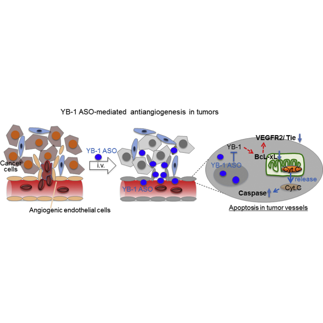
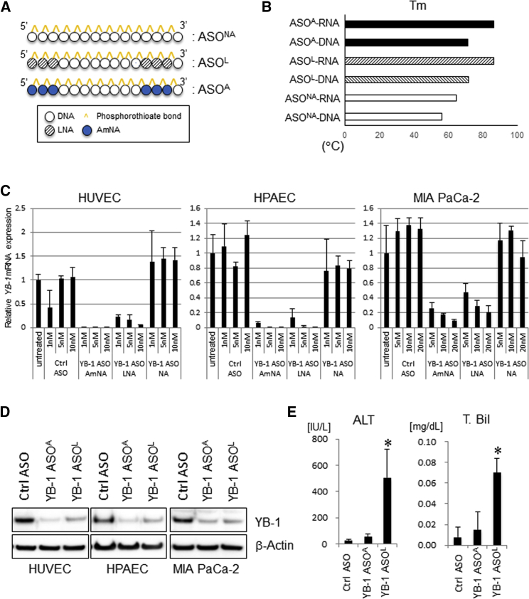
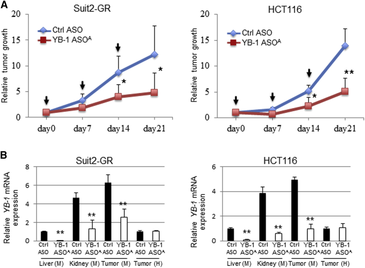
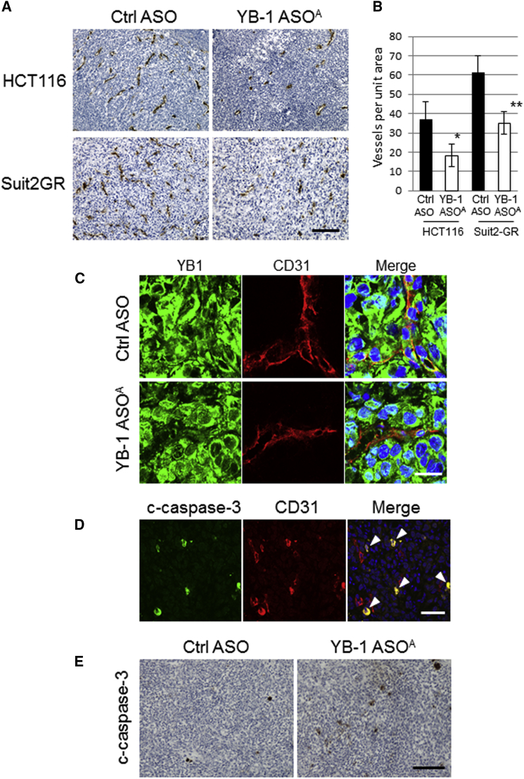
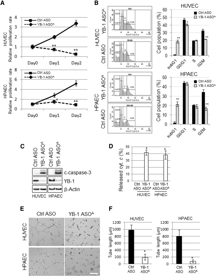
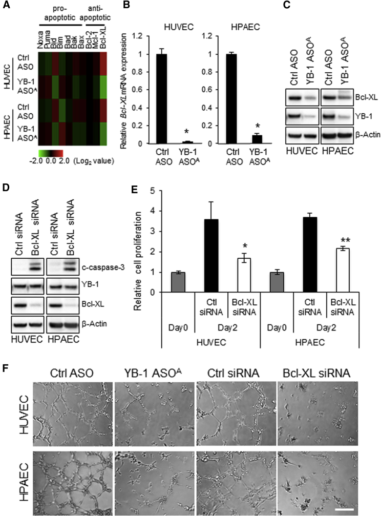
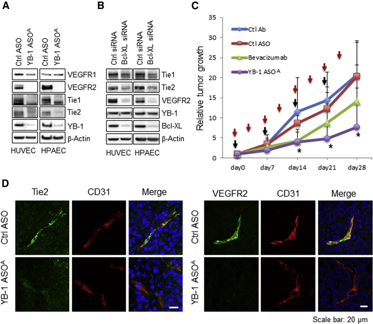
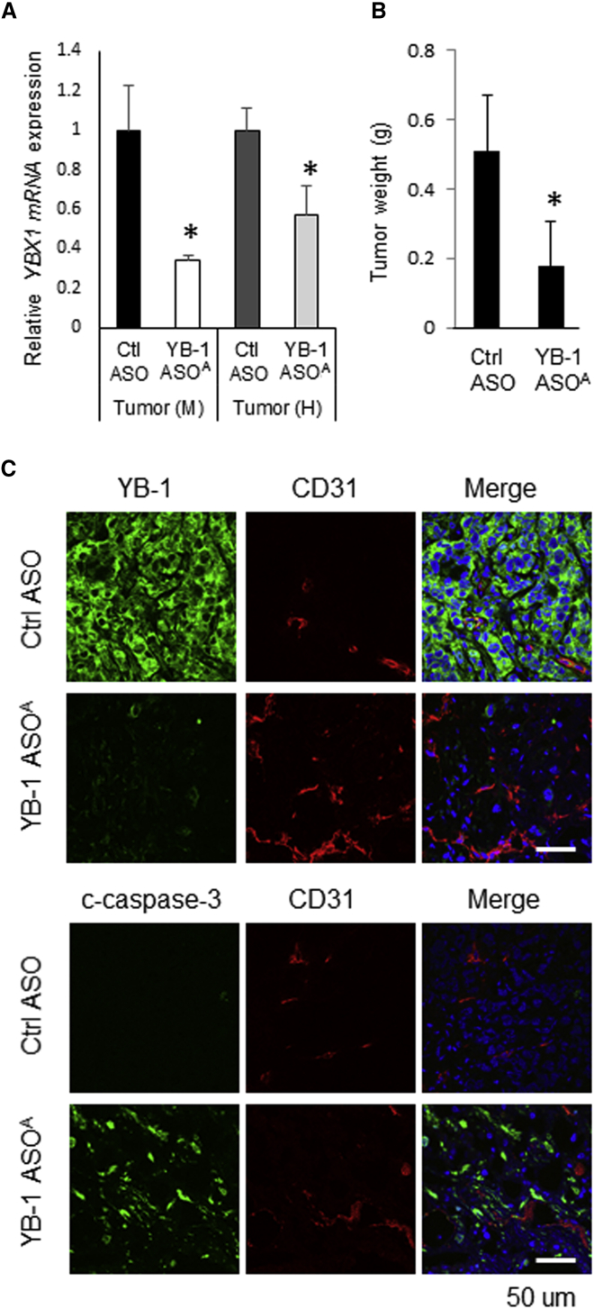
Y-box binding protein-1 (YB-1), involved in cancer progression and chemoradiation resistance, is overexpressed in not only cancer cells but also tumor blood vessels. In this study, we investigated the potential value of amido-bridged nucleic acid (AmNA)-modified antisense oligonucleotides (ASOs) targeting YB-1 (YB-1 ASOA) as an antiangiogenic cancer therapy. YB-1 ASOA was superior to natural DNA-based ASO or locked nucleic acid (LNA)-modified YB-1 ASO in both knockdown efficiency and safety, the latter assessed by liver function. YB-1 ASOA administered i.v. significantly inhibited YB-1 expression in CD31-positive angiogenic endothelial cells, but not in cancer cells, in the tumors. With regard to the mechanism of its antiangiogenic effects, YB-1 ASOA downregulated both Bcl-xL/VEGFR2 and Bcl-xL/Tie signal axes, which are key regulators of angiogenesis, and induced apoptosis in vascular endothelial cells. In the xenograft tumor model that had low sensitivity to anti-VEGF antibody, YB-1 ASOA significantly suppressed tumor growth; not only VEGFR2 but also Tie2 expression was decreased in tumor vessels. In conclusion, YB-1/Bcl-xL/VEGFR2 and YB-1/Bcl-xL/Tie signal axes play pivotal roles in tumor angiogenesis, and YB-1 ASOA may be feasible as an antiangiogenic therapy for solid tumors.

Keywords: ASO; Tie; VEGFR2; YB-1; tumor angiogenesis.


Figures

None
Graphical abstract
Figure 1
Figure 1
Construction of YB-1 ASOs and Their Effects on YB-1 mRNA and Protein Expression In Vitro (A) Schematic structures of several types of YB-1 ASOs. (B) Tm values of YB-1 ASO duplexes with DNA and RNA target strands. (C) qRT-PCR of human YBX1 mRNA in HUVECs, HPAECs, and MIA PaCa-2 cells transfected with YB-1 ASOs or control ASO (normalized to B2M). n = 3. (D) Immunoblots for YB-1 protein in HUVECs, HPAECs, and MIA PaCa-2 cells transfected with YB-1 ASOs or control ASO (5 nM). β-actin is the loading control. (E) Effects of i.v. administered YB-1 ASOA, YB-1 ASOL, or control ASO (once per week for 3 weeks at 10 mg/kg body weight) on liver function, evaluated by alanine aminotransferase (ALT) and T. Bil values. *p < 0.01; n = 4 per group. Data are means ± SD.
Figure 2
Figure 2
Antitumor Efficacy of i.v. Administered YB-1 ASOA in Mice Harboring Subcutaneous Tumor Xenografts (A) Antitumor efficacy of i.v. administered YB-1 ASOA (once per week for 3 weeks at 10 mg/kg body weight) in mice bearing HCT116 or Suit2-GR xenografts. *p < 0.05, **p < 0.01; n = 7 per group. Black arrows represent administered time points of ASOs. (B) qRT-PCR of mouse YBX1 mRNA in liver, kidney, or tumor and human YBX1 mRNA in tumor tissues from mice harboring HCT116 and Suit2-GR xenografts and receiving YB-1 ASOA treatments (once per week for 3 weeks at 10 mg/kg body weight). **p < 0.001; n = 7 per group. Values are normalized to those for mouse or human 18S rRNAs. Data are means ± SD.
Figure 3
Figure 3
i.v. Administered YB-1 ASOA Suppresses Tumor Growth In Vivo through Antiangiogenic Effects on Host Vascular Cells (A) Representative immunostaining images showing CD31 in HCT116 and Suit2-GR tumors after YB-1 ASOA treatment (once per week for 3 weeks at 10 mg/kg body weight). Scale bar, 200 μm. (B) Quantification of microvascular density, assessed by CD31 staining per field, in HCT116 and Suit2-GR tumor tissues. *p < 0.05, **p < 0.01; n = 6. Data are means ± SD. (C) Representative immunostaining images for YB-1 and CD31 in Suit2-GR tumor tissues in mice treated with YB-1 ASOA or control ASO (once per week for 3 weeks at 10 mg/kg body weight). Scale bar, 20 μm. (D) Representative immunostaining images showing cleaved caspase-3 (c-caspase-3) and mouse CD31 in Suit2-GR tumor tissues from mice receiving i.v. YB-1 ASOA treatment (once per week for 3 weeks at 10 mg/kg body weight). Yellow signal (arrowhead) in merged images represents apoptosis (c-caspase-3) in CD31-positive angiogenic endothelial cells. Scale bar, 50 μm. The fluorescent signal images (C and D) were obtained using immunofluorescence laser confocal microscopy. (E) Immunostaining of c-caspase-3 in Suit2-GR tumors from mice treated with i.v. administered YB-1 ASOA (once per week for 3 weeks at 10 mg/kg body weight). YB-1 ASOA-treated, but not control, tumors had apoptotic cells. Scale bar, 200 μm.
Figure 4
Figure 4
Involvement of YB-1 in Proliferation, Apoptosis, and Tube Formation in Vascular Endothelial Cells HUVECs and HPAECs were transfected with YB-1 ASOA or control ASOA (5 nM) and subjected to the following analyses. (A) Cell proliferation analysis with the WST-8 assay. *p < 0.05, **p < 0.01; n = 3. (B) Cell-cycle distribution, analyzed in HUVEC and HPAEC by staining the DNA content with propidium iodide. The distribution was measured by flow cytometry. *p < 0.05, **p < 0.01; n = 3. (C) Western blot analysis shows robustly increased c-caspase-3 (apoptosis) and decreased YB-1 levels in YB-1 ASOA-transfected cells. β-actin is the loading control. (D) Cytochrome c release from the IMS was detected in HUVECs and HPAECs transfected with YB-1 ASOA in the presence of a pan-caspase inhibitor, Z-VAD-FMK, under immunofluorescence laser confocal microscopy. Data were obtained from 50 cells in each of three independent experiments. Quantitated levels of released cytochrome c were higher in cells with YB-1 ASOA transfection than in controls. *p < 0.001. (E) Representative images showing tube formation in HUVEC and HPAEC cultures transfected with YB-1 ASOA or control. Scale bar, 200 μm. (F) Quantitation of total tube length in 10 randomly chosen images using ImageJ software. *p < 0.001. Data are means ± SD.
Figure 5
Figure 5
YB-1 ASOA-Induced Apoptosis and Inhibition of Tube Formation Are Mediated by Bcl-xL Reduction in Vascular Endothelial Cells (A) Heatmap analysis of cDNA microarray data showing highly downregulated Bcl-xL expression, among Bcl-2 family genes, in HUVECs and HPAECs transfected with YB-1 ASOA (5 nM). (B) Downregulation of Bcl-xL expression was confirmed by qRT-PCR analysis (normalized to values for 18S). *p < 0.01; n = 3. Data are means ± SD. (C) Immunoblots for Bcl-xL and YB-1 in HUVECs and HPAECs transfected with YB-1 ASOA or control ASO (5 nM). β-actin is the loading control. (D) Western blot analysis shows an increase in c-caspase-3 but no change in YB-1 protein levels in HUVECs and HPAECs transfected with Bcl-xL siRNA (5 nM). (E) Cell proliferation, assessed by WST-8 assay, was decreased in HUVECs and HPAECs by transfection with Bcl-xL siRNA. *p < 0.05, **p < 0.01. (F) Capillary-like tube formation was inhibited by transfection with Bcl-xL siRNA or YB-1 ASOA in HUVECs and HPAECs. Scale bar, 200 μm.
Figure 6
Figure 6
Knockdown of YB-1/Bcl-xL Downregulates VEGFR2 and Tie-1/-2 Expression in Angiogenic Endothelial Cells in Tumor Microenvironments (A and B) Western blot analysis showing inhibition of VEGFR2 and Tie-1/-2 expression in HUVECs or HPAECs transfected with YB-1 ASOA (5 nM) (A) or Bcl-xL siRNA (20 nM) (B). β-actin is the loading control. (C) Antitumor efficacy of YB-1 ASOA treatment (once per week for 4 weeks at 10 mg/kg body weight) was superior to that of bevacizumab (twice per week for 4 weeks at 10 mg/kg body weight) in mice bearing Suit2-GR xenografts. *p < 0.05; n = 9. Data are means ± SD. Red and black arrows represent administered time points of bevacizumab and ASOs, respectively. (D) Immunohistochemistry for VEGFR2, Tie2, and CD31 in the Suit2-GR tumors using laser confocal microscopy. The images show inhibition of Tie2 signals in CD31-positive angiogenic endothelial cells in tumor tissues treated with i.v. administered YB-1 ASOA (once per week for 4 weeks at 10 mg/kg body weight), but not in the controls. Scale bar, 20 μm.
Figure 7
Figure 7
Antitumor Efficacy of i.p. Administered YB-1 ASOA in Mice Harboring Peritoneal Dissemination (A) qRT-PCR analysis shows an inhibition of mouse and human YBX1 expression in peritoneally disseminated RMG-1 tumors treated with i.p. administered YB-1 ASOA. *p < 0.05; n = 3. Values are normalized to those for mouse and human 18S rRNAs. (B) Disseminated RMG-1 tumor growth was inhibited on day 21 in mice treated with YB-1 ASOA (once per week for 3 weeks at 10 mg/kg body weight) compared with control ASOs. *p < 0.05; n = 5. Data are means ± SD. (C) Immunohistochemistry for CD31 and YB-1 or c-caspase-3 using laser confocal microscopy. The images show decreased YB-1 and increased c-caspase-3 levels in not only CD31-positive angiogenic endothelial cells but also most disseminated RMG-1 cancer cells after i.p. administration of YB-1 ASOA. Scale bar, 50 μm.

Similar articles

Cited by

References

    1. Crooke S.T. Antisense strategies. Curr. Mol. Med. 2004;4:465–487. - PubMed
    1. Wu H., Lima W.F., Zhang H., Fan A., Sun H., Crooke S.T. Determination of the role of the human RNase H1 in the pharmacology of DNA-like antisense drugs. J. Biol. Chem. 2004;279:17181–17189. - PubMed
    1. Galarneau A., Min K.L., Mangos M.M., Damha M.J. Assay for evaluating ribonuclease H-mediated degradation of RNA-antisense oligonucleotide duplexes. Methods Mol. Biol. 2005;288:65–80. - PubMed
    1. Vester B., Wengel J. LNA (locked nucleic acid): high-affinity targeting of complementary RNA and DNA. Biochemistry. 2004;43:13233–13241. - PubMed
    1. Wahlestedt C., Salmi P., Good L., Kela J., Johnsson T., Hökfelt T., Broberger C., Porreca F., Lai J., Ren K. Potent and nontoxic antisense oligonucleotides containing locked nucleic acids. Proc. Natl. Acad. Sci. USA. 2000;97:5633–5638. - PMC - PubMed