. 2018 May;138(5):1052-1061.
doi: 10.1016/j.jid.2017.11.033. Epub 2017 Dec 12.

DLX3-Dependent STAT3 Signaling in Keratinocytes Regulates Skin Immune Homeostasis

Affiliations

DLX3-Dependent STAT3 Signaling in Keratinocytes Regulates Skin Immune Homeostasis

Shreya Bhattacharya et al. J Invest Dermatol. 2018 May.

Abstract

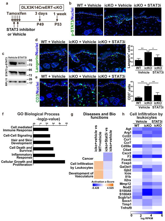
Epidermal-specific deletion of the homeobox transcription regulator DLX3 disrupts keratinocyte differentiation and results in an IL-17-linked psoriasis-like skin inflammation. To identify the epidermal initiating signals produced by DLX3-null keratinocytes, we performed acute deletion of DLX3 in adult epidermis using a tamoxifen-inducible Krt14-cre/ERT system. K14CreERT;DLX3fl/fl skin exhibited dysregulated expression of differentiation-associated genes, upregulation of proinflammatory cytokines, and accumulation of Langerhans cells and macrophages within 3 days of tamoxifen-induced DLX3 ablation. We also observed increased accumulation of IL-17A-secreting Vγ4 γδ T cells and heightened levels of IL-17 and IL-36 family of cytokines starting 1 week after DLX3 deletion. Interestingly, transcriptome profiling of K14CreERT;DLX3fl/fl epidermis at 3 days identified activated STAT3 as a transcriptional regulator and revealed differential expression of STAT3 signaling-related genes. Furthermore, activation of STAT3 was strongly increased in K14CreERT;DLX3fl/fl skin, and topical treatment with an inhibitor of STAT3 activation attenuated the immune phenotype. RNA-seq analysis of vehicle and STAT3 inhibitor treated K14CreERT;DLX3fl/fl skin identified differentially expressed genes associated with inhibition of leukocyte infiltration. Collectively, our results show that DLX3 is a critical regulator of STAT3 signaling network that maintains skin homeostasis.


Conflict of interest statement

Conflict of Interest

Dr. Mark Udey has a perceived conflict of interest because he is the Editor of the Journal of Investigative Dermatology. He addressed this by recusing himself from all aspects of the review and editorial processes related to this submission. All other authors state no conflict of interest.

Figures

Figure 1
Figure 1. Acute epidermal deletion of DLX3 in adult skin induces altered keratinocyte differentiation and barrier defects
(a) Schematic representation of topical tamoxifen treatment and sample collection post-tamoxifen (TAM) treatment. (b) RNA Sequencing tracks for the DLX3 locus shows distinct peaks for three exons in WT (DLX3fl/fl) and absence of peaks at exon 2 and 3 in icKO (K14-CreERT2;Dlx3fl/fl) confirming conditional inducible deletion of DLX3. (c) Log2 fold changes between DLX3 WT and icKO groups were used for Gene Ontology (GO) terms. The graph summarizes all the biological processes detected by GO enrichment analysis at 3 days, 1 week and 2 weeks in the epidermal transcriptome. Antilog of p-values was used for graphical representation. (p <0.05). (d) Heatmap showing differential expression of epidermal differentiation complex genes (EDC) at 3 days and 1 week. Values represent normalized log2 RPKM (n = 3 per group). (e) Immunohistochemistry of skin sections with antibodies for Keratin 6 (K6) and differentiation marker Filaggrin at 3 days and 1 week after TAM treatment. All sections were counterstained with Hoechst to establish landmarks and visualize tissue morphology and nuclei. The white dotted line demarcates epidermis from dermis. Scale bar = 100um
Figure 2
Figure 2. Inducible DLX3 deletion in keratinocytes triggers proinflammatory cytokine expression and infiltration of T cells, primarily IL-17A-secreting T-cells
(a) Heatmaps displaying cytokines upregulated and downregulated in epidermis and dermis 3 days after TAM treatment. (b) Upregulation of subsets of cytokines and chemokines in epidermis and dermis 1 week after DLX3 deletion by TAM treatment. (c) Immunofluorescent labeling of Langerhans cells using langerin antibody. Bottom panel, Bar graph depicting significant changes in total numbers of Langerhans cells in icKO epidermis in comparison to WT. (d) Representative images of immunofluorescence staining shows increases in CD3+ T cells in icKO versus WT skin at 3 days and 1 week after DLX3-ablation. Bar graph indicates numbers of CD3+ T cells in WT and icKO skin. (e) Immunolabeling of CD4+ T cells subpopulation in WT and icKO skin at 3 days and 1 week. Hoechst was used as a counterstain. Graph represents numbers of CD4 + cells in the dermal compartment. The white dotted line demarcates epidermis from dermis. Scale bar = 100um (f) Flow cytometry analysis characterized the total and specific subpopulations of IL-17A secreting T cells at 3 days. (g) Increased total number of IL- 17A producing cells and their sub-populations 1 week after TAM treatment. p-value significance *>0.05 and **>0.01.
Figure 3
Figure 3. Activation of STAT3 signaling in DLX3 deleted icKO skin
(a) Upstream regulators of 3 days gene dataset by IPA (left panel) and the differentially observed genes related to the potential upstream regulator of STAT3 activation (right panel). (b) Immunofluorescence for phosphorylated STAT3 (p-STAT3) showed increased detection in 3 days and 1 week icKO epidermis post-TAM treatment. Percentage of positive p-STAT3 cells in the epidermis are presented in the bar graph. The white dotted line indicates epidermal-dermal boundary. Scale bar = 100um (c) Western blot analysis of p-STAT3 and total STAT3. RPL11 was used as an internal control for normalization. Quantitative analysis of western blot results showed increased expression of p-STAT3 with respect to total STAT3 in icKO skin at 3 days and 1 week.
Figure 4
Figure 4. Treatment with STAT3 specific inhibitor cryptotanshinone reduced inflammation in icKO skin
(a) Schematic representation of TAM and cryptotanshinone (STAT3i) or Vehicle treatment and subsequent sample collection after treatment. (b) Immunolabeling of p-STAT3 in vehicle and STAT3i treated skin. (c) Western blot analysis showed reduction of p-STAT3 expression after STAT3i treatment in icKO skin. (d) Langerin staining and quantitation of Langerin-positive Langerhans cells in icKO skin after vehicle and STAT3i treatment. WT with vehicle treatment was used as control. (e) Immunofluoresecence with CD3 antibody of vehicle and STAT3i treated WT and icKO skin. Bar graph demonstrates decrease of CD3+ T cells after STAT3i treatment. p-value significance *>0.05 and **>0.01. (f) GO functional classification of differentially expressed genes. (g) Z-score identifies the activation or inhibition of biological functions. (h) Gene expression in icKO vehicle and cryptotanshinone treated samples. Color key: red represents upregulated and green downregulated expression.

References

    1. Akitsu A, Ishigame H, Kakuta S, Chung SH, Ikeda S, Shimizu K, et al. IL-1 receptor antagonist-deficient mice develop autoimmune arthritis due to intrinsic activation of IL-17-producing CCR2(+)Vgamma6(+)gammadelta T cells. Nature communications. 2015;6:7464. - PMC - PubMed
    1. Albanesi C, De Pita O, Girolomoni G. Resident skin cells in psoriasis: a special look at the pathogenetic functions of keratinocytes. Clinics in dermatology. 2007;25(6):581–8. - PubMed
    1. Augustin I, Gross J, Baumann D, Korn C, Kerr G, Grigoryan T, et al. Loss of epidermal Evi/Wls results in a phenotype resembling psoriasiform dermatitis. The Journal of experimental medicine. 2013;210(9):1761–77. - PMC - PubMed
    1. Blumberg H, Dinh H, Trueblood ES, Pretorius J, Kugler D, Weng N, et al. Opposing activities of two novel members of the IL-1 ligand family regulate skin inflammation. The Journal of experimental medicine. 2007;204(11):2603–14. - PMC - PubMed
    1. Botchkarev VA. Integration of the Transcription Factor-Regulated and Epigenetic Mechanisms in the Control of Keratinocyte Differentiation. The journal of investigative dermatology Symposium proceedings /the Society for Investigative Dermatology, Inc [and] European Society for Dermatological Research. 2015;17(2):30–2. - PMC - PubMed

Publication types