. 2018 Mar;176(3):1981-1992.
doi: 10.1104/pp.17.01374. Epub 2017 Dec 15.

LRX Proteins Play a Crucial Role in Pollen Grain and Pollen Tube Cell Wall Development

Affiliations

LRX Proteins Play a Crucial Role in Pollen Grain and Pollen Tube Cell Wall Development

Tohnyui Ndinyanka Fabrice et al. Plant Physiol. 2018 Mar.

Abstract

Leu-rich repeat extensins (LRXs) are chimeric proteins containing an N-terminal Leu-rich repeat (LRR) and a C-terminal extensin domain. LRXs are involved in cell wall formation in vegetative tissues and required for plant growth. However, the nature of their role in these cellular processes remains to be elucidated. Here, we used a combination of molecular techniques, light microscopy, and transmission electron microscopy to characterize mutants of pollen-expressed LRXs in Arabidopsis (Arabidopsisthaliana). Mutations in multiple pollen-expressed lrx genes cause severe defects in pollen germination and pollen tube growth, resulting in a reduced seed set. Physiological experiments demonstrate that manipulating Ca2+ availability partially suppresses the pollen tube growth defects, suggesting that LRX proteins influence Ca2+-related processes. Furthermore, we show that LRX protein localizes to the cell wall, and its LRR-domain (which likely mediates protein-protein interactions) is associated with the plasma membrane. Mechanical analyses by cellular force microscopy and finite element method-based modeling revealed significant changes in the material properties of the cell wall and the fine-tuning of cellular biophysical parameters in the mutants compared to the wild type. The results indicate that LRX proteins might play a role in cell wall-plasma membrane communication, influencing cell wall formation and cellular mechanics.


Figures

Figure 1.
Figure 1.
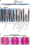
Seed set and pollen viability. A, Schematic representation of the LRX proteins indicating the site of Citrine insertion. B, Representative images of fully developed siliques of the wild type and different single, double, triple, and quadruple lrx mutants as well as complemented lines. Most severe defects are observed in the triple and quadruple mutants. Seed set is partially or fully restored in complemented lines (mutant background and gene used for complementation are separated by a double colon). C, Alexander staining of anthers showing comparable pollen viability in the wild type and lrx8/9 and lrx8/9/11 mutants.
Figure 2.
Figure 2.
Pollen germination and PT growth. A, Percentage germinated PGs, burst grains, and burst tubes after 2 h of incubation in vitro, showing higher proportion of burst PGs and PTs in the lrx mutants. Percentage of burst PTs is based on the germinated PTs. Nongerminated PGs were distinguished from germinated PGs by the absence of a detectable PT. B, Transmission electron micrographs of transverse sections through stigma/papilla surface, stigmatic papillae, and transmitting tract of the ovary. While wild-type PGs germinate and PTs grow, lrx8/9/11 mutant grains mostly burst, discharge their content into the stigmatic papillar matrix (spm), and shrink. Eventually, compared to the wild type, fewer lrx8/9/11 mutant PTs (some indicated by arrows) grow through the papillar apoplast into the ovary transmitting tract (tt) that is surrounded by septum cells (sc). C, Typical wild-type PTs (mostly regular cylindrical) and lrx PTs (bulging, bursting, budding) phenotypes are shown. tt, transmitting tract; sc, septum cells. Bars = 10 µm in B and 20 µm in C.
Figure 3.
Figure 3.
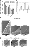
PT growth in different Ca2+ regimes. A, Average length of PTs in standard (std) PGM (containing 5 mm CaCl2), PGM containing reduced [Ca2+] (2 mm CaCl2), and in PGM supplemented with 5 µm and 15 µm LaCl3. While lower [Ca2+] or LaCl3 treatments reduced PT growth in the wild type, these regimes improved PT growth in lrx8/9 and lrx8/9/11 mutants (n ≥ 200; error bar = se; different letters indicate significant differences, t test, P < 0.05). B, The percentage change in PT length under different Ca2+ regimes compared to their corresponding values in standard PGM. The highest positive effect is obtained with the lrx8/9/11 mutant at 5 µM LaCl3. C, Kymographs showing continuous and intermittent growth of wild-type and lrx PTs, respectively, in standard PGM. D, In PGM containing 5 µm LaCl3, continuous growth is slightly perturbed in the wild type, while intermittent growth is partially restored to continuous growth in lrx mutants.
Figure 4.
Figure 4.
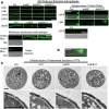
Immunolabeling and ultrastructural analyses of pollen tube cell walls. A, The labeling of ER/Golgi- synthesized cell wall components (with LM2, LM6 LM19, LM15, and JIM20) and plasma membrane synthesized cell wall components (with aniline blue). The labeling for ER/Golgi-synthesized wall components is significantly weaker in lrx8/9, lrx8/9/11, and lrx8/9/10 compared to the wild type. The labeling for plasma membrane-synthesized callose (aniline blue) in mutants is stronger than in the wild type and also found at the tip. Even longer exposure of wild-type PTs demonstrates callose labeling in the shank but not at the tip. Xyloglucanase treated PTs still show significantly lower labeling for pectin (LM20, LM6) compared to the wild type, and no labeling for xyloglucan (LM15). DIC captures are shown for aniline-stained wild-type PT and LM15-labeled PT treated by xyloglucanase to show position of the PTs. B, Cytoplasmic content released from the PT strongly stain for cell wall components, as shown here for pectin (LM6). C, TEM transverse sections of PTs. The mutant PT outer cell walls are more loose/fibrous and the inner wall thicker, reflecting the higher accumulation of callose (strongest in lrx8/9/11) compared to the wild type. Bars = 20 µm in A, 10 µm in B, and 1 µm in C.
Figure 5.
Figure 5.
Visualization and quantification of intracellular Ca2+ dynamics. Time series of YC 3.60 fluorescence showing a [Ca2+] gradient with a tip-localized increase in wild-type (A) and lrx8/9/11 (B) PTs (top). A strong increase in [Ca2+] is seen in the mutant prior to bursting. Graphs show fluorescence of Ca2+-unbound YC 3.60 (blue line), Ca2+-bound YC 3.60 (red line), and the ratio representing the Ca2+ signal (green line) with the spike in the mutant prior to bursting.
Figure 6.
Figure 6.
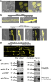
LRX-Citrine and LRR-Citrine localization. A, LRR11-Citrine fluorescence in PGs. B, LRX11-Citrine localization in cell wall and cytoplasm in turgid and plasmolyzed PTs. C and D, LRR11-Citrine (C) localizes to the cell wall-plasma membrane and cytoplasm in turgid PTs, but retracts with the plasma membrane and cytoplasm in the plasmolyzed cells, as does LRR4-Citrine (D) in hypocotyl cells of pLRX4::LRR4-Citrine transgenic seedlings. Bar = 10 µm. (E) Western blot of total extracts (left) and membrane fractions (right) of wild-type and pLRX4::LRR4-Citrine transgenic (T) seedlings probed with an anti-GFP, anti-LHC1a, or anti-FBP antibody to detect LRR4-Citrine, the membrane protein LHC1a, and the cytoplasmic protein FBP, respectively. Tobacco leaf material expressing LRR11-Citrine (T) and nontransgenic tobacco (WT) was purified in the same way and LRR11-Citrine also copurified with the membrane fraction. Bars = 10 µm in A to D.
Figure 7.
Figure 7.
Biophysical properties of PTs deduced by FEM-based modeling. Compared to the wild type, turgor pressure (light-gray box) is significantly increased in the lrx8/9 and lrx8/9/11. The stiffness of the cell wall (dark-gray box) is significantly increased in the lrx8/9/11 mutant compared to the wild type, which is similar in lrx8/9. Statistics: t test, *P < 0.03, **P < 0.0001, n ≥ 50. In addition, the box plots show considerable skewness of turgor and cell wall stiffness in lrx mutants as revealed by the larger difference between the median (line) and the mean (stroked line) as well as the range of the whiskers compared to the wild type where the deviations are smaller. Given the skewness, the mean values shown are calculated from the log normalized data.

Comment in

References

    1. Alexander MP. (1969) Differential staining of aborted and nonaborted pollen. Stain Technol 44: 117–122 - PubMed
    1. Alonso JM, Stepanova AN, Leisse TJ, Kim CJ, Chen H, Shinn P, Stevenson DK, Zimmerman J, Barajas P, Cheuk R, et al. (2003) Genome-wide insertional mutagenesis of Arabidopsis thaliana. Science 301: 653–657 - PubMed
    1. Bai L, Zhang G, Zhou Y, Zhang Z, Wang W, Du Y, Wu Z, Song CP (2009) Plasma membrane-associated proline-rich extensin-like receptor kinase 4, a novel regulator of Ca signalling, is required for abscisic acid responses in Arabidopsis thaliana. Plant J 60: 314–327 - PubMed
    1. Baluška F, Šamaj J, Wojtaszek P, Volkmann D, Menzel D (2003) Cytoskeleton-plasma membrane-cell wall continuum in plants. Emerging links revisited. Plant Physiol 133: 482–491 - PMC - PubMed
    1. Battey NH, James NC, Greenland AJ, Brownlee C (1999) Exocytosis and endocytosis. Plant Cell 11: 643–660 - PMC - PubMed

Publication types

MeSH terms